Bilder
Videos
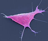
13633140 - Human induced pluripotent cell, SEM
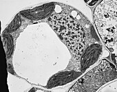
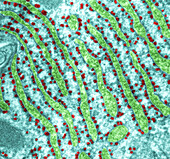
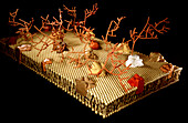
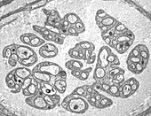
13618624 - Timothy grass mesophyll cell, TEM
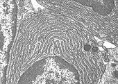
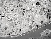
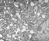
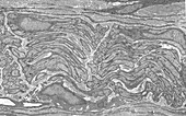
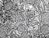
13613414 - Rough endoplasmic reticulum, TEM
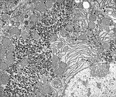
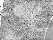
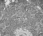
13613409 - Liver cell, TEM
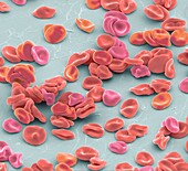
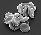
13585683 - Red blood cells, SEM
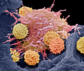
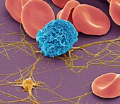
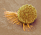
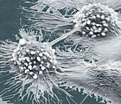
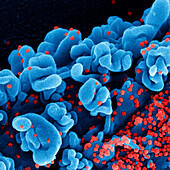
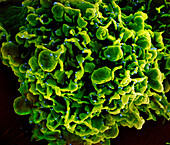
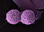
14078715 - (CAR) T-cell therapy, SEM
13756283 - Organoid, SEM
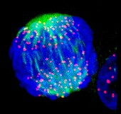
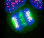
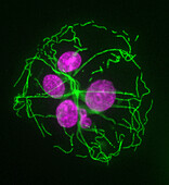
13755887 - Mitosis, light micrograph
13755807 - Mitosis, light micrograph

13755805 - Daughter cells after mitosis, light micrograph
13755274 - Daughter cells after mitosis, light micrograph
13672044 - Cervical cancer cell, TEM
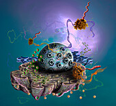
13632782 - Cell interior, illustration
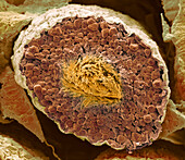
13620794 - Testis, TEM
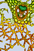
13620313 - Stinging nettle root, light micrograph
13620302 - Marram grass, light micrograph
13620301 - Marram grass, light micrograph
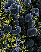
13585662 - Blood cells, SEM
13755945 - Rough endoplasmic reticulum, TEM
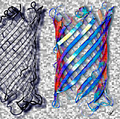
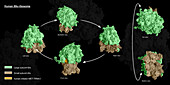
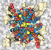
13685854 - Bacterial outer membrane protein, illustration
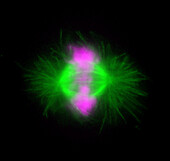
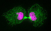
13672555 - Human cell in anaphase, light micrograph
13672547 - Human cell in interphase, light micrograph
13633074 - Cell membrane, illustration

13632841 - Cellular protein transport, illustration
13613430 - Spermatogonia, TEM

13613416 - Liver, TEM

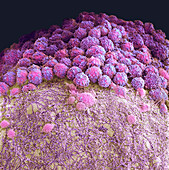
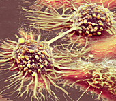
14077653 - Cervical cancer cell, SEM
14077650 - Cervical cancer cell, SEM
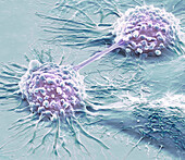
14077626 - Dividing HeLa cells, SEM

13755840 - Mitosis, light micrograph
13755551 - Mitosis, light micrograph
13754736 - Mitosis in binucleate cells, light micrograph
13743093 - Organoid, SEM.
13672833 - Human 80s ribosome, illustration
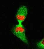
13672554 - Human cell in telophase, light micrograph
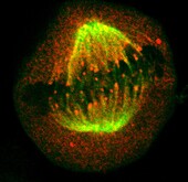
13672552 - Human cell in metaphase, light micrograph
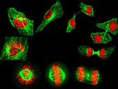
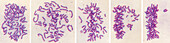
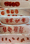
13672549 - Human cells showing the stages of cell division, light micrograph
13672535 - Cell nucleus in prometaphase, light micrograph
13634177 - Testis, SEM

13613422 - Pancreas, TEM
13613399 - Protein synthesis, TEM

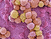
13613370 - Lung cancer cells dividing, SEM
13613362 - Oral squamous cell carcinoma, SEM
13755097 - Mitosis, light micrograph
13754706 - Mitosis, light micrograph
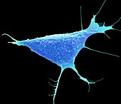
13686219 - Stem cell-derived neurons, light micrograph
13672557 - Two human cells in interphase, light micrograph
13672533 - Human cell in anaphase, light micrograph
13620792 - Ovary, TEM
13620292 - Soft rush stem, light micrograph
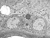
13613420 - Seminiferous epithelium, TEM
13613408 - Liver, TEM
13524737 - Stem cell technology
13755895 - Interphase cell treated with Taxol, light micrograph
13755889 - Mitosis, light micrograph
13755804 - Mitosis, light micrograph
13754738 - Mitosis in binucleate cells, light micrograph
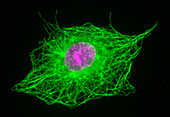
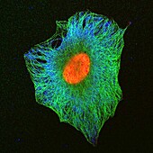
13672566 - Human fibroblast cells, light micrograph
13672331 - Eumycetoma infected tissue sample, light micrograph
13672324 - Cell infected by SARS-CoV-2 Omicron virus particles, SEM
13634166 - Testis, SEM
13613413 - Insulin granules, TEM
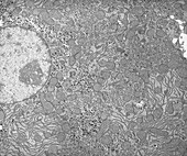
13613405 - Liver, TEM
13435529 - Cell infected by SARS-CoV-2 virus particles, SEM
12395231 - Red blood cells, SEM
13754741 - Mitosis in tetranucleate cells, light micrograph
13686117 - Sodium potassium channel, illustration
13672556 - Human cell in early prophase, light micrograph
13647381 - Protein synthesis, illustration
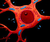
13632840 - Nerve cell, illustration
13620790 - Nerve, TEM
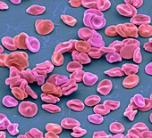
13585669 - Red blood cells, SEM
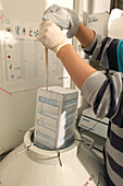
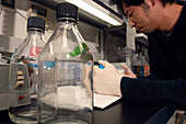
13524733 - Induced pluripotent stem cells being frozen
14077627 - Dividing HeLa cells, SEM
13755896 - Interphase cell treated with Colchicine, light micrograph
13755891 - Cytokinesis, light micrograph

13754742 - Mitosis in tetranucleate cells, light micrograph

13733278 - Animal cell structure, illustration

13672561 - Human cell in metaphase, light micrograph
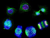
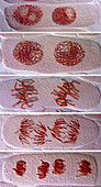
13672553 - Stages of cell division, light micrograph
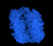
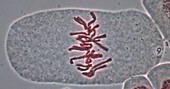
13672532 - Human chromosomes in early anaphase, light micrograph
13633134 - Human induced pluripotent cell
13613421 - Nerve, TEM
13613369 - Lung cancer cells dividing, SEM
13525474 - Gap junction, TEM
14077617 - Dividing HeLa cells, SEM
13755886 - Mitosis, light micrograph
13754700 - Mitosis, light micrograph
13672828 - Human 80s ribosome, illustration
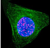
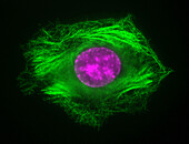
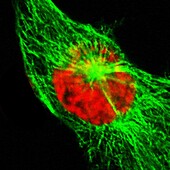
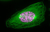
13672534 - Human cell showing nucleus and tubulin, light micrograph
13634182 - Testis, SEM

13620785 - Microvilli, TEM
13613428 - Epididymis, TEM
13613407 - Microvilli, TEM
13613398 - Sertoli cell, TEM
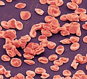
13585660 - Red blood cells, SEM
13435526 - Cell infected by SARS-CoV-2 virus particles, SEM

13416490 - Cells infected by Covid-19 virus particles, SEM

13405953 - Microtubule, computer model
nächste Seite
Zellbilogie Bilder ❘ Science Photo Library