Seminiferous epithelium, TEM
Bildnummer 13613420
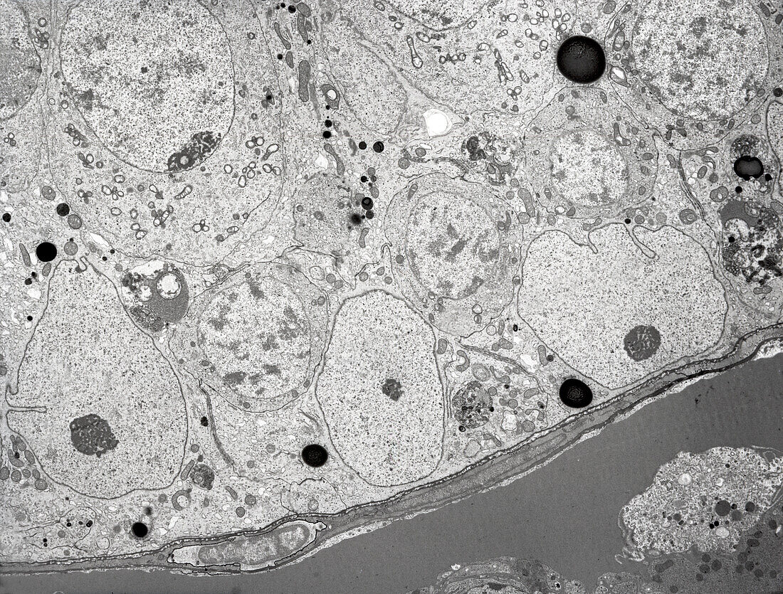
| Transmission electron micrograph (TEM) of the ultrastructure of the basal region of the seminiferous epithelium of the testis. Three Sertoli cell nuclei with nucleoli lie close to the basal lamina. Tight junctions occur between adjacent Sertoli cells which serve as the blood-testis barrier. Above and between the Sertoli cells are three zygotene primary spermatocytes displaying round nuclei with condensed chromosomes. Part of a large pachytene primary spermatocyte is seen at upper left. Magnification: x2, 000 when printed at a width of 10cm. | |
| Lizenzart: | Lizenzpflichtig |
| Credit: | Science Photo Library / Microscape |
| Bildgröße: | 4724 px × 3584 px |
| Modell-Rechte: | nicht erforderlich |
| Eigentums-Rechte: | nicht erforderlich |
| Restrictions: | - |
Preise für dieses Bild ab 15 €
Universitäten & Organisationen
(Informationsmaterial Digital, Informationsmaterial Print, Lehrmaterial Digital etc.)
ab 15 €
Redaktionell
(Bücher, Bücher: Sach- und Fachliteratur, Digitale Medien (redaktionell) etc.)
ab 30 €
Werbung
(Anzeigen, Aussenwerbung, Digitale Medien, Fernsehwerbung, Karten, Werbemittel, Zeitschriften etc.)
ab 55 €
Handelsprodukte
(bedruckte Textilie, Kalender, Postkarte, Grußkarte, Verpackung etc.)
ab 75 €
Pauschalpreise
Rechtepakete für die unbeschränkte Bildnutzung in Print oder Online
ab 495 €