Sertoli cell, TEM
Bildnummer 13613398
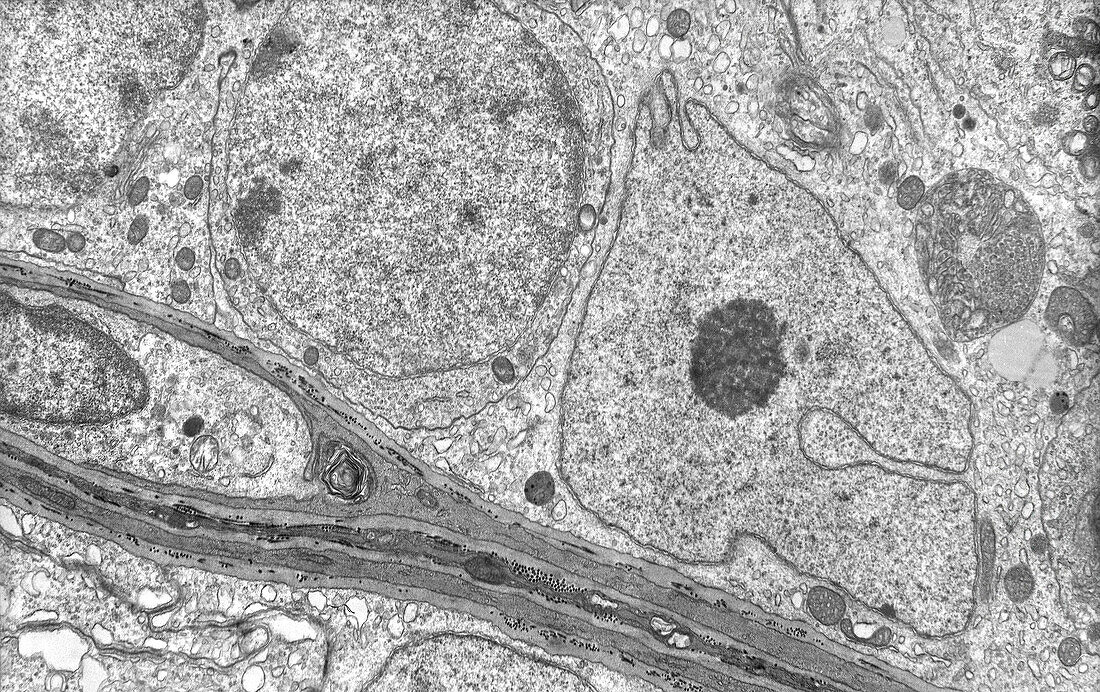
| Transmission electron micrograph (TEM) of the ultrastructure of a Sertoli cell to the right and a circular preleptotene primary spermatocyte to the left. The basal lamina of the seminiferous epithelium is at bottom. Magnification: x7, 000 when printed at a width of 10cm. | |
| Lizenzart: | Lizenzpflichtig |
| Credit: | Science Photo Library / Microscape |
| Bildgröße: | 5291 px × 3329 px |
| Modell-Rechte: | nicht erforderlich |
| Eigentums-Rechte: | nicht erforderlich |
| Restrictions: | - |
Preise für dieses Bild ab 15 €
Universitäten & Organisationen
(Informationsmaterial Digital, Informationsmaterial Print, Lehrmaterial Digital etc.)
ab 15 €
Redaktionell
(Bücher, Bücher: Sach- und Fachliteratur, Digitale Medien (redaktionell) etc.)
ab 30 €
Werbung
(Anzeigen, Aussenwerbung, Digitale Medien, Fernsehwerbung, Karten, Werbemittel, Zeitschriften etc.)
ab 55 €
Handelsprodukte
(bedruckte Textilie, Kalender, Postkarte, Grußkarte, Verpackung etc.)
ab 75 €
Pauschalpreise
Rechtepakete für die unbeschränkte Bildnutzung in Print oder Online
ab 495 €
Keywords
- Basallamina,
- biologisch,
- Einfarbig,
- Elektronenmikroskopie,
- elektronenmikroskopische Aufnahme,
- Hoden,
- männliche Fortpflanzung,
- männliches Fortpflanzungssystem,
- Niemand,
- schwarz und weiß,
- Sertoli-Zelle,
- tem,
- transmissionselektronenmikroskopische Aufnahme,
- Ultrastruktur,
- Zellbilogie,
- Zytologie,
- Zytologisch