Mitosis, light micrograph
Bildnummer 13755840
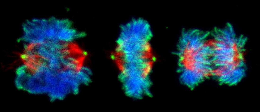
| Fluorescent light micrograph of cells during mitosis (nuclear division). Mitosis is the formation of two daughter nuclei from one parent nucleus. Fluorescent markers have been used to highlight DNA (deoxyribonucleic acid, blue), alpha tubulin (red), a component of microtubules, and topoisomerase II (green), an enzyme that plays a role in the condensation of chromosomes prior to mitosis. The cell at left is in prometaphase with condensed chromosomes. The cell at centre is in metaphase, where the chromosomes align along the centre of the cell. The chromosomes start to move to the opposite poles, guided by microtubules, during anaphase (right). | |
| Lizenzart: | Lizenzpflichtig |
| Credit: | Science Photo Library / DR. JUAN F. GIMENEZ-ABIAN |
| Bildgröße: | 4534 px × 1953 px |
| Modell-Rechte: | nicht erforderlich |
| Eigentums-Rechte: | nicht erforderlich |
| Restrictions: | - |
Preise für dieses Bild ab 15 €
Universitäten & Organisationen
(Informationsmaterial Digital, Informationsmaterial Print, Lehrmaterial Digital etc.)
ab 15 €
Redaktionell
(Bücher, Bücher: Sach- und Fachliteratur, Digitale Medien (redaktionell) etc.)
ab 30 €
Werbung
(Anzeigen, Aussenwerbung, Digitale Medien, Fernsehwerbung, Karten, Werbemittel, Zeitschriften etc.)
ab 55 €
Handelsprodukte
(bedruckte Textilie, Kalender, Postkarte, Grußkarte, Verpackung etc.)
ab 75 €
Pauschalpreise
Rechtepakete für die unbeschränkte Bildnutzung in Print oder Online
ab 495 €
Keywords
- alpha-Tubulin,
- Biologie,
- biologisch,
- Chromosom,
- Chromosomen,
- Desoxiribonukleinsäure,
- DNA,
- Einteilung,
- Enzym,
- Fluoreszenz,
- fluoreszierend,
- Genetik,
- kopierend,
- lichtmikroskopische Aufnahme,
- Mikroskopie,
- Mitose,
- mitotische Spindel,
- Niemand,
- Prometaphase,
- Reproduktion,
- schwarzer Hintergrund,
- Separieren,
- Teilen,
- Wachstum,
- Zellbilogie,
- Zelle,
- Zellen,
- Zytologie,
- Zytologisch,
- Zytoskelett