Human cell in telophase, light micrograph
Bildnummer 13672554
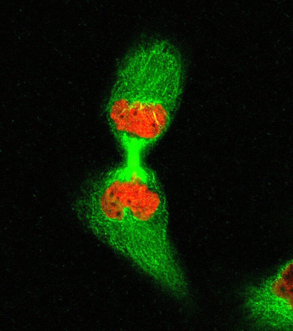
| HeLa cell in telophase, fluorescence light micrograph. Chromatin is stained red and the microtubules forming the spindle are stained green. The chromosomes have separated and decondensed, and the midbody has formed. | |
| Lizenzart: | Lizenzpflichtig |
| Credit: | Science Photo Library / DR MATTHEW DANIELS |
| Bildgröße: | 3050 px × 3439 px |
| Modell-Rechte: | nicht erforderlich |
| Eigentums-Rechte: | nicht erforderlich |
| Restrictions: | - |
Preise für dieses Bild ab 15 €
Universitäten & Organisationen
(Informationsmaterial Digital, Informationsmaterial Print, Lehrmaterial Digital etc.)
ab 15 €
Redaktionell
(Bücher, Bücher: Sach- und Fachliteratur, Digitale Medien (redaktionell) etc.)
ab 30 €
Werbung
(Anzeigen, Aussenwerbung, Digitale Medien, Fernsehwerbung, Karten, Werbemittel, Zeitschriften etc.)
ab 55 €
Handelsprodukte
(bedruckte Textilie, Kalender, Postkarte, Grußkarte, Verpackung etc.)
ab 75 €
Pauschalpreise
Rechtepakete für die unbeschränkte Bildnutzung in Print oder Online
ab 495 €