Bilder
Videos
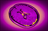
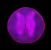
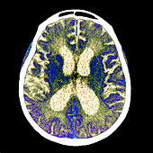
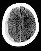
13736254 - Healthy brain, CT scan
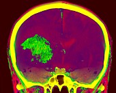
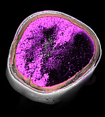
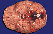
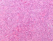
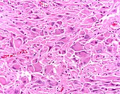
13733436 - Glioblastoma

13673066 - Structure of the eye, illustration
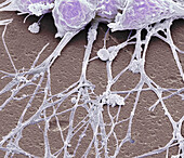
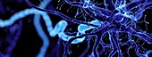
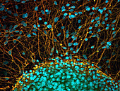
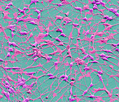
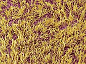
13671968 - Neurones, SEM
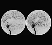
13600400 - Treatment for clotting of the middle cerebral artery
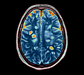
13599520 - Multiple sclerosis, MRI scan
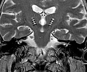
13452125 - Parkinson's disease electrode implants, MRI scan
13386572 - Intraparenchymal haemorrhage, CT scan

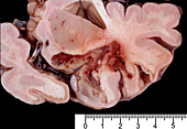
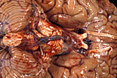
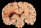
13357902 - Human brain abscess
13357892 - Human Brain, Subarachnoid Basal Hemorrhage

13357877 - Human Brain, Hydrocephalus
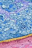
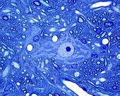
13357185 - Myenteric nerve plexus, TEM
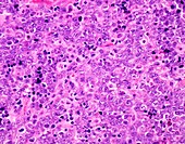
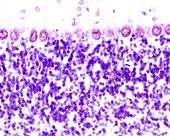
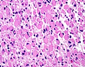
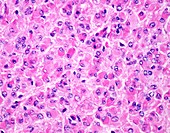
13297607 - Diffuse large B-cell lymphoma, light micrograph
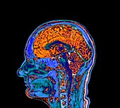
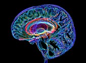
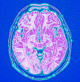
13296512 - Healthy brain, MRI scan
13296461 - Haemangioblastoma, light micrograph
13296454 - Anaplastic ependymoma, light micrograph
13296447 - Choroid plexus papilloma, light micrograph

13296443 - Choroid plexus carcinoma, light micrograph
13258325 - Visual pathways, illustration

13257842 - Brain reward pathway, illustration
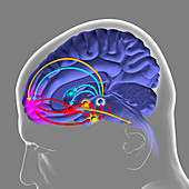
13257833 - Brain reward pathway, illustration
13257814 - Brain reward pathway, illustration
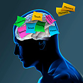
13245521 - Memory loss, conceptual illustration
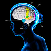
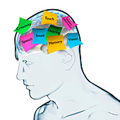
13245499 - Areas of the brain, conceptual illustration

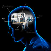
13224891 - AI, illustration
13223523 - Nerve cell, illustration
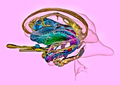
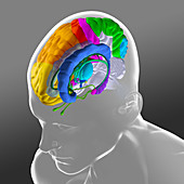
12967551 - Human brain and limbic system,3D MRI-based image

14077877 - Augusta Dejerine-Klumpke and Joseph Jules Dejerine, neurologists
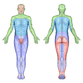
14077605 - Dermatome skin sensory areas, illustration

13952070 - Human brain, MRI scan
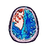
13951872 - Hydrocephalus, 3D MRI scan
13951693 - Shingles rash
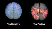
13951492 - Healthy brain and brain with tau aggregations, MRI scans

13765756 - Fahr syndrome, CT scan
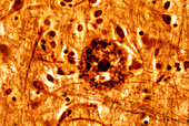
13756138 - Senile plaque in cerebral cortex, light micrograph
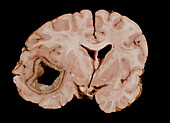
13733395 - Human brain abscess
13732600 - Nerve structures of the retina, 1894 illustration
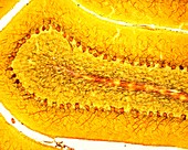
13686369 - Spinal cord, light micrograph
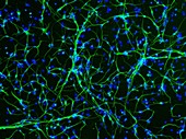
13686222 - Stem cell-derived neurons, light micrograph
13673777 - Ischaemic stroke, CT scan
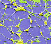
13671966 - Neurones, SEM
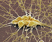
13633181 - Pluripotent derived neurones, SEM
13633137 - Pluripotent derived neurones, SEM
13599645 - Alzheimer's disease, CT scan
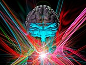
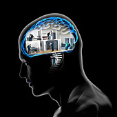
13505291 - Brain activity, conceptual illustration
13426620 - Microglia, light micrograph
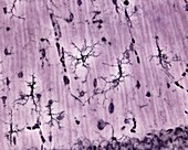
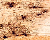
13416597 - Cerebellar cortex Purkinje cells, light micrograph

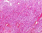
13387286 - Meningioma, light micrograph
13357875 - Human brain. Subarachnoid hemorrage
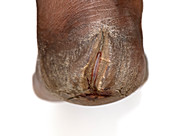
13356973 - Foot affected by leprosy
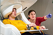
13297883 - Patient in a MEG scanner
13297620 - Fibrous meningioma, light micrograph
13296428 - Gemistocytic astrocytoma, light micrograph
13272265 - Brain lining, SEM
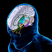
13258356 - Brain anatomy, illustration
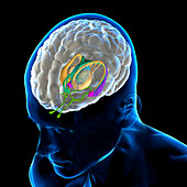
13258335 - Brain anatomy, illustration
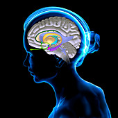
13258316 - Brain anatomy, illustration
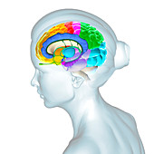
13258308 - Brain anatomy, illustration
13257841 - Brain reward pathway, illustration

13257738 - Sensory areas of the brain, illustration
13245497 - Dementia, conceptual illustration
13223548 - Brain scan, conceptual illustration
12987480 - Cerebellar cortex, light micrograph
12987473 - Cerebellar cortex, light micrograph
12987455 - Fibrous astrocytes, light micrograph
12967556 - Deep brain stimulation,3D CT-based image
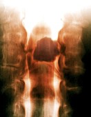
12960795 - Spinal cord tumour,X-ray myelography image
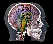
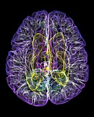
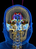
12960771 - Human skull and brain,CT and MRI scans
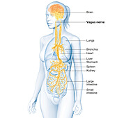
14078555 - Active brain and energetic vagus nerve, illustration
13952071 - Human brain, MRI scan
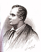
13951843 - Charles Bell, Scottish anatomist, illustration

13951695 - Shingles rash
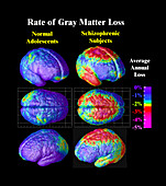
13951045 - Grey matter loss in very early-onset schizophrenia, brain maps
13838686 - Type I amyloid-beta filaments, illustration
13756142 - Senile plaque in cerebral cortex, light micrograph
13647476 - Neurons, SEM

13599648 - Alzheimer's disease, CT scan
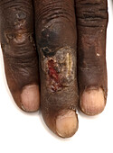
13453706 - Ulcer on finger of leprosy patient
13452060 - Subdural hematoma, CT scan
13445668 - Limbic system in Alzheimer's disease, 3D MRI scan

13426633 - Oligodendroglia, light micrograph
13426618 - Neuron cell body, light micrograph

13426606 - Pacinian corpuscle in periosteum, light micrograph
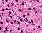
13416604 - Astrocytes, light micrograph

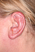
13405792 - Bell's palsy
13387801 - Brain hippocampus neurons, fluorescence light micrograph
13386986 - Glioblastoma brain cancer, CT scan
13357885 - Human Brain, Hemorrhagic Infarct
13297632 - Oligodendroglioma, light micrograph
13297631 - Meningothelial meningioma, light micrograph
13297622 - Meningothelial meningioma, light micrograph
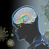
13297403 - Loss of smell in Covid-19, illustration
13296439 - Subependymal giant cell astrocytoma, light micrograph
13296429 - Gemistocytic astrocytoma, light micrograph

13258338 - Brain anatomy, illustration
13258329 - Brain anatomy, illustration
13257739 - Sensory areas of the brain, illustration
13245517 - Areas of the brain, conceptual illustration
13245501 - Areas of the brain, conceptual illustration
nächste Seite
Neurologie Bilder ❘ Science Photo Library