Patient in a MEG scanner
Bildnummer 13297883
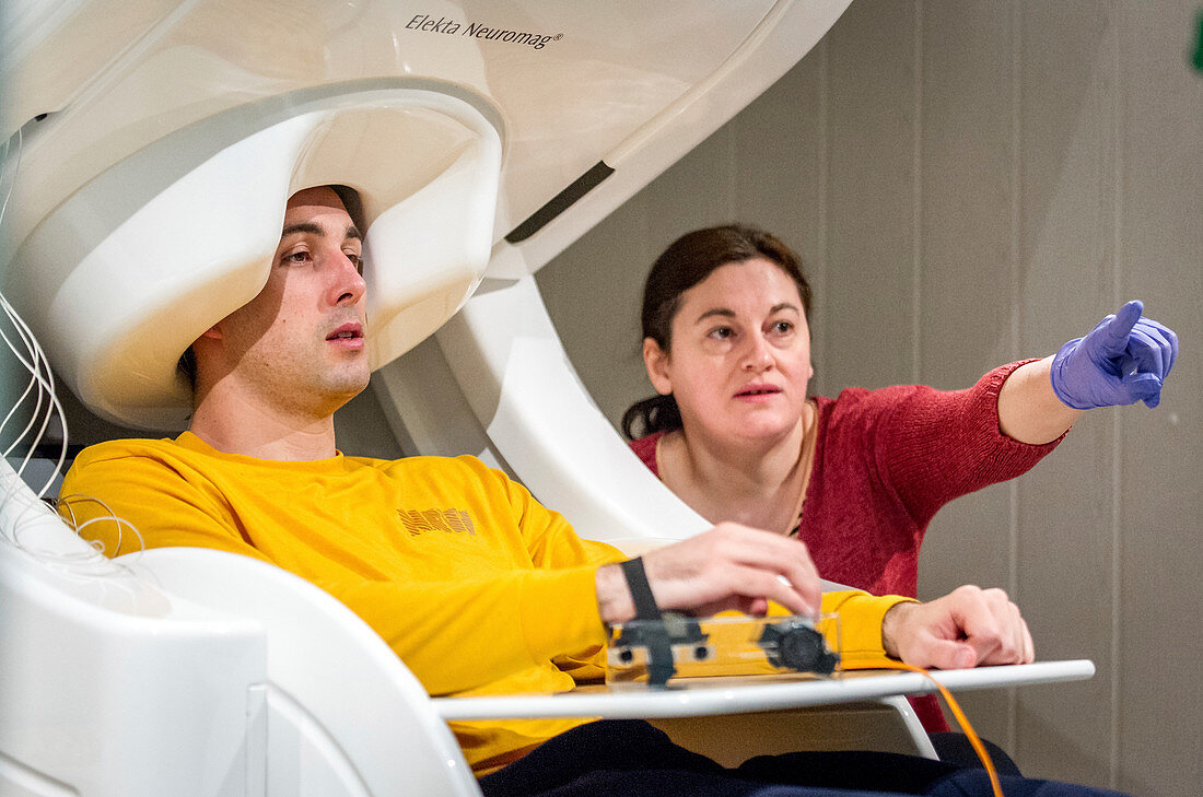
| Patient in a magnetoencephalograph (MEG) scanner. A researcher directing a patient to perform various tasks as a MEG scanner maps brain activity. MEG is a functional neuroimaging technique which maps brain activity by recording magnetic fields produced by the electrical currents naturally occurring in the brain. Unlike electroencephalography (EEG) scanners, these typically use highly sensitive SQUIDs (Superconducting Quantum Interference Devices) to detect the extremely small magnetic fields and associated gradients generated by neural activity in different parts of the brain. By measuring the minute changes in magnetic fields generated by the cerebral cortex while resting, while thinking and while engaged in some particular task, deficits in the brain can be measured and localized. The resulting data may be used to investigate psychiatric and neurological disorders such as ADHD, epilepsy and age-related memory and cognitive disorders such as Alzheimer's disease and dementia. Photographed at the National Intrepid Center of Excellence (NICoE) at the Walter Reed National Military Medical Center, Bethesda, Maryland, USA, on 21 March 2017. | |
| Lizenzart: | Lizenzpflichtig |
| Credit: | Science Photo Library / US AIR FORCE / J.M. Eddins Jr.l |
| Bildgröße: | 3683 px × 2442 px |
| Modell-Rechte: | Derzeit liegt noch kein Release vor. Bitte kontaktieren Sie uns vor Verwendung. |
| Eigentums-Rechte: | nicht erforderlich |
| Restrictions: |
|
Preise für dieses Bild ab 15 €
Universitäten & Organisationen
(Informationsmaterial Digital, Informationsmaterial Print, Lehrmaterial Digital etc.)
ab 15 €
Redaktionell
(Bücher, Bücher: Sach- und Fachliteratur, Digitale Medien (redaktionell) etc.)
ab 30 €
Werbung
(Anzeigen, Aussenwerbung, Digitale Medien, Fernsehwerbung, Karten, Werbemittel, Zeitschriften etc.)
ab 55 €
Handelsprodukte
(bedruckte Textilie, Kalender, Postkarte, Grußkarte, Verpackung etc.)
ab 75 €
Pauschalpreise
Rechtepakete für die unbeschränkte Bildnutzung in Print oder Online
ab 495 €
Keywords
- 2 Leute,
- 21. Jahrhundert,
- Amerikanisch,
- Analyse,
- Ausrüstung,
- Behandlung,
- Bild,
- Bildgebung,
- biomedizinisch,
- Diagnose,
- Forscher,
- Forschung,
- Frau,
- Funktional,
- Gehirn,
- Gehirnaktivität,
- Gesundheitswesen,
- Hirnscan,
- Magnet,
- Magnetfeld,
- Magnetismus,
- Mann,
- Männlich,
- Maryland,
- Medizin,
- medizinisch,
- Medizinische Technologie,
- Menschen,
- neural,
- Neuroimaging,
- Neurologie,
- neurologisch,
- neuromagnetisch,
- neuronale Aktivität,
- Neurowissenschaften,
- Nicoe,
- Performance,
- Person,
- Psychiatrie,
- Psychologie,
- Scan,
- Technik,
- Testen,
- Tintenfisch,
- uns,
- USA,
- Vereinigte Staaten,
- Weiblich,
- Zentrum