Choroid plexus papilloma, light micrograph
Bildnummer 13296447
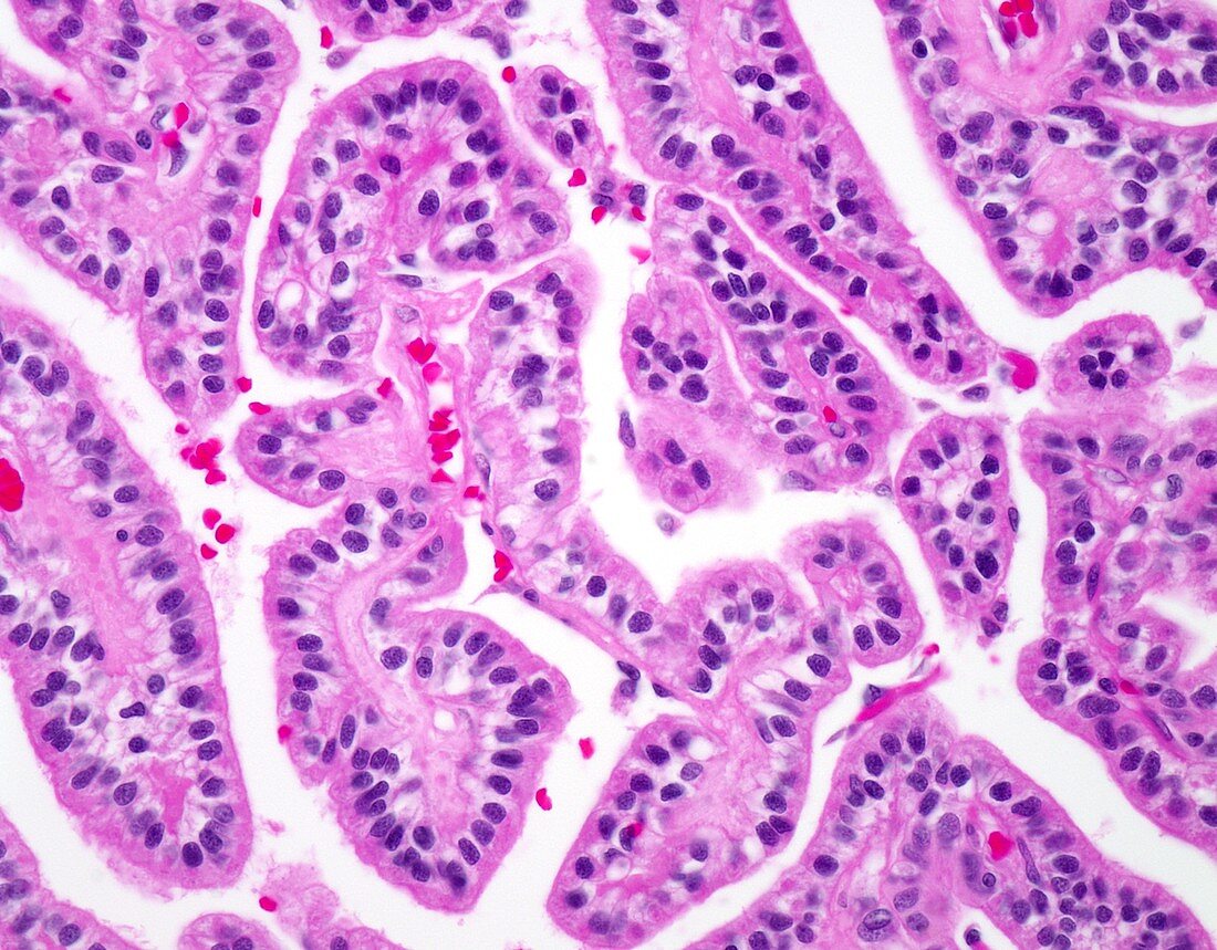
| Choroid plexus papilloma, light micrograph. Choroid plexus tumours comprise less than 1% of all brain tumours. Almost 80% of cases occur in children making up 2% to 4% of pediatric brain tumours. The most common location is lateral ventricles. Given their location, the presenting symptoms and signs are related to hydrocephalus and increased intracranial pressure. Choroid plexus papillomas are circumscribed, papillary masses consisting of pseudostratified epithelial cells lining fibrovascular cores. Note the orderly arrangement of epithelial cells around fibrovascular cores. Cytologic atypia is absent and mitotic activity is not increased. Choroid plexus papillomas are low-grade tumours (WHO Grade 1) and cured by total resection (100% 5-yr survival). | |
| Lizenzart: | Lizenzpflichtig |
| Credit: | Science Photo Library / WEBPATHOLOGY |
| Bildgröße: | 4096 px × 3200 px |
| Modell-Rechte: | nicht erforderlich |
| Eigentums-Rechte: | nicht erforderlich |
| Restrictions: | - |
Preise für dieses Bild ab 15 €
Universitäten & Organisationen
(Informationsmaterial Digital, Informationsmaterial Print, Lehrmaterial Digital etc.)
ab 15 €
Redaktionell
(Bücher, Bücher: Sach- und Fachliteratur, Digitale Medien (redaktionell) etc.)
ab 30 €
Werbung
(Anzeigen, Aussenwerbung, Digitale Medien, Fernsehwerbung, Karten, Werbemittel, Zeitschriften etc.)
ab 55 €
Handelsprodukte
(bedruckte Textilie, Kalender, Postkarte, Grußkarte, Verpackung etc.)
ab 75 €
Pauschalpreise
Rechtepakete für die unbeschränkte Bildnutzung in Print oder Online
ab 495 €
Keywords
- abnormal,
- Epilepsie,
- Gehirn,
- Histologie,
- histologisch,
- Histopathologie,
- histopathologisch,
- Karzinom,
- Kondition,
- Krankheit,
- Krebs,
- krebsartig,
- Lichtmikroskop,
- lichtmikroskopische Aufnahme,
- maligne,
- Malignom,
- Neurologie,
- neurologisch,
- Niemand,
- Onkologie,
- pädiatrisch,
- Papillom,
- Pathologie,
- pathologisch,
- Störung,
- Tumor,
- ungesund,
- zentrales Nervensystem