Bilder
Videos
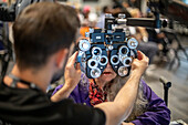
13954624 - Woman picking out frames for glasses
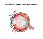
13736725 - Retinal detachment, illustration
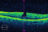
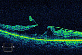
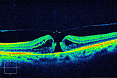
13687022 - Macular pucker with lamellar hole, OCT scan
13687021 - Macular pucker with lamellar hole, OCT scan
13674391 - Liquid gel eye drops
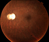
13613554 - Intraocular lens subluxation, fundoscopy
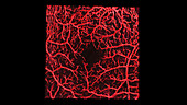
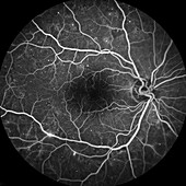
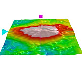
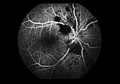
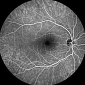
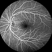
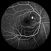
13951663 - Retinal blood vessels, OCT angiography scan
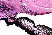
13756214 - Schlemm canal in eye, light micrograph
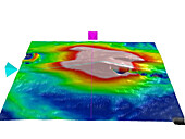
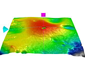
13733185 - Diabetic macular oedema, OCT macular cube scan

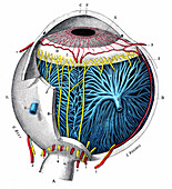
13732601 - Nerve structures of the retina, 1894 illustration
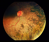
13613586 - Retinal vasculitis, fundoscopy
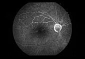
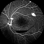
13613568 - Retina damage from diabetes, angiogram

13524567 - Slit lamp eye examination
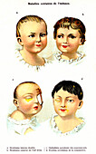
13951800 - Infant eye conditions, illustration

13613650 - Retina damage from diabetes, fundoscopy
13524552 - Slit lamp eye examination

13954623 - People having eye tests
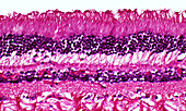
13950466 - Human retina cells, light micrograph

13736724 - Retinal detachment, illustration
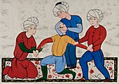
13736082 - Eye surgery, 17th century illustration
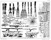
13732604 - Nerve structures of the retina, 1894 illustration
13633364 - Cat eye with hypertension, illustration
13613640 - Retina damage from diabetes, angiogram
13613577 - Retinal vasculitis, fundoscopy
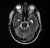
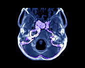
13599593 - Retinal detachment, MRI scan
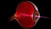
13585921 - Light entering human eye, illustration

13525322 - Severe burning of an eye from gas poisoning, illustration
13733188 - Macular pucker with lamellar hole, OCT macular cube scan
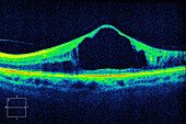
13613558 - Vitreomacular traction, OCT scan
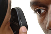
13523862 - Eye examination
13954622 - Woman having eyes tested
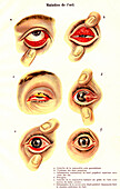
13951804 - Eye diseases, illustration
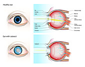
13736718 - Normal eye and eye with cataract, illustration

13736716 - Healthy eye and diabetic retinopathy, illustration
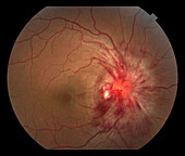
13613564 - Papilloedema, fundoscopy
13599517 - Pulsatile exophthalmos, CT angiogram

13585905 - Light entering human eye, illustration
13524540 - Eye test
13756213 - Choroid and retina, light micrograph
13733186 - Diabetic macular oedema, OCT macular cube scan
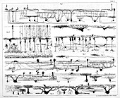
13732603 - Nerve structures of the retina, 1894 illustration
13732016 - Anterior ischemic optic neuropathy, angiogram
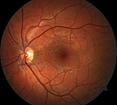
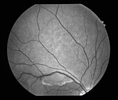
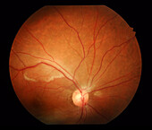
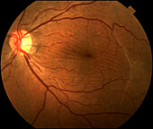
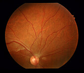
13613550 - Healthy eye, fundoscopy
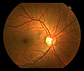
13613548 - Healthy eye, fundoscopy
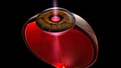
13585907 - Light entering human eye, illustration

13954579 - Hidrocystoma on a woman's eye
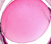
13756200 - Eye lens fibres, light micrograph
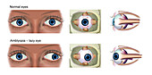
13736714 - Normal eye and amblyopia, illustration
13732018 - Temporal epiretinal membranes, fundoscopy
13613634 - Pigmentary retinopathy, fundoscopy
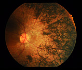
13613632 - Pigmentary retinopathy, fundoscopy
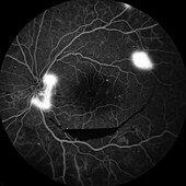
13613546 - Healthy eye, angiogram

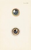
12969886 - Kayser-Fleischer rings in eyes due to liver disease

14077544 - Glaucoma eye drops
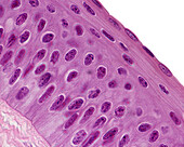
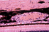
13756183 - Human cornea, light micrograph
13732020 - Hemispheric retinal artery occlusion, angiogram
13687019 - Diabetic macular oedema, OCT scan
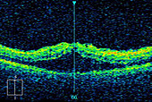
13687015 - Diabetic macular oedema, OCT scan
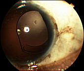
13613652 - Intraocular lens subluxation

13613545 - Healthy eye, angiogram
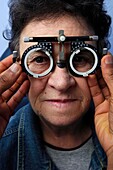
13524861 - Eyesight test trial frames
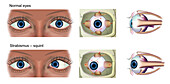
13736727 - Normal eye and strabismus, illustration

13687016 - Diabetic macular oedema, OCT scan
13613581 - Retinal vasculitis, fundoscopy
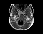
13599516 - Pulsatile exophthalmos, CT angiogram
13524558 - Eye test chart
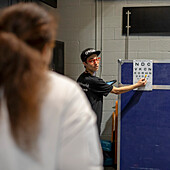
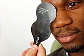
13954618 - Eye test with Snellen chart
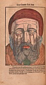
13736205 - Eye surgery, 16th century illustration

13733184 - Diabetic macular oedema, OCT macular cube scan

13736715 - Healthy eye and diabetic retinopathy, illustration
13733178 - Eye anatomy, illustration
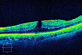
13687020 - Lamellar macular hole, OCT scan
13613547 - Healthy eye, angiogram
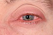
12644137 - Anterior uveitis

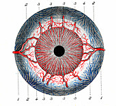
13736135 - Eye blood vessels, illustration
13733177 - Eye anatomy, illustration
13732017 - Central serous retinopathy, angiogram

13613584 - Retinal vasculitis, angiogram
13613570 - Retina damage from diabetes, angiogram

12490410 - Amsler grid used to detect Macular Degeneration
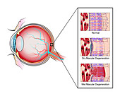
13736723 - Macular degeneration, illustration

13736722 - Macular degeneration, illustration

13613647 - Retina damage from diabetes, angiogram
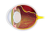
13585904 - Human eye anatomy, illustration
13525317 - Cornea in acute stage of burning, illustration
13524554 - Eye examination
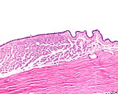
13426612 - Conjunctiva and sclera, light micrograph

13377650 - Long-sightedness corrected, illustration
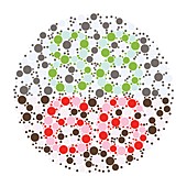
13356702 - Colour blindness test chart, illustration
13356680 - Colour blindness test chart, illustration

13356676 - Colour blindness test chart, illustration
13356662 - Colour blindness test chart, illustration

13356652 - Colour blindness test chart, illustration
13356640 - Colour blindness test chart, illustration
13356542 - Colour blindness test chart, illustration

13356485 - Colour blindness test chart, illustration
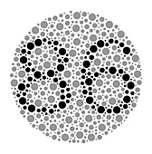
13356455 - Colour blindness test chart, illustration
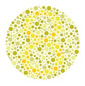
13356424 - Colour blindness test chart, illustration

13356413 - Colour blindness test chart, illustration
nächste Seite
Ophthalmologie Bilder ❘ Science Photo Library