Bilder
Videos
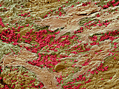
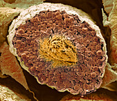
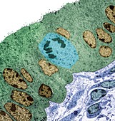
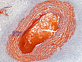
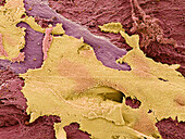
13839160 - Uterus during menstruation, SEM
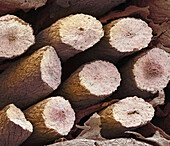
13634177 - Testis, SEM
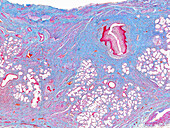
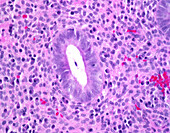
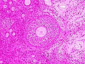
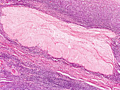
13619784 - Human Bartholin's gland, light micrograph
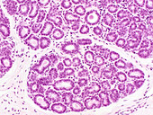
13619694 - Human mammary gland in pregnancy, light micrograph
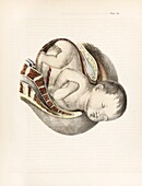
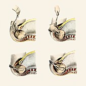
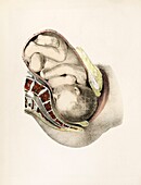
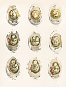
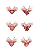
13525044 - Foetus in breech position, 19th century illustration
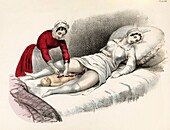
13525017 - Childbirth, 19th century illustration
13525014 - Childbirth, 19th century illustration
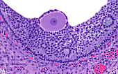
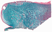
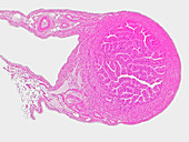
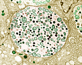
13950537 - Tertiary ovarian follicle, light micrograph

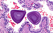
13950434 - Corpus amylaceous, light micrograph
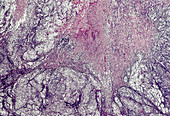
13756573 - Chronic endometritis, light micrograph
13619765 - Human uterus, light micrograph
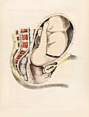
13525362 - Uterus during pregnancy, 19th century illustration
13950437 - Corpus luteum cells, light micrograph
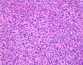
13756578 - Uterine leiomyoma, light micrograph
13733289 - Ovarian and uterine cycles, illustration
13685534 - Prostatitis, illustration
13673496 - Human testicle, light micrograph
13634166 - Testis, SEM
13619761 - Human uterus, light micrograph
13619698 - Inactive human mammary gland, light micrograph
13619697 - Human urethra, light micrograph
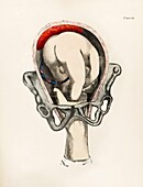
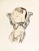
13525314 - Forceps delivery, 19th century illustration
13525311 - Forceps delivery, 19th century illustration
13525020 - Foetus in breech position, 19th century illustration
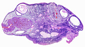
13950446 - Testis epididymis, light micrograph
13685532 - Prostatitis, illustration
13619767 - Human uterus, light micrograph
13619751 - Human placenta, light micrograph
13619744 - Human umbilical cord, light micrograph
13619715 - Monkey ovary, light micrograph
13619682 - Rabbit ovary, light micrograph
13950432 - Corpora amylacea, light micrograph
13756593 - Serous carcinoma, light micrograph
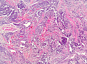
13756563 - Adenosarcoma of uterus, light micrograph

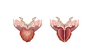
13685531 - Healthy and cancerous prostate glands, illustration
13619745 - Human uterus, light micrograph
13619739 - Human uterus, light micrograph
13619728 - Human placenta, light micrograph
13619702 - Monkey ovary, light micrograph
13756586 - Uterine leiomyosarcoma, light micrograph
13756576 - Epithelioid trophoblastic tumour, light micrograph
13634182 - Testis, SEM
13619757 - Human uterus, light micrograph
13619749 - Human umbilical cord, light micrograph
13619426 - Sperm, TEM
13452721 - Tubal ligation clips, X-ray
13444121 - Penile cancer, light micrograph

13435562 - Ovum, illustration
13377737 - Adenomyosis, light micrograph
13950440 - Uterus decidua cells, light micrograph
13950433 - Corpus amylaceous, light micrograph
13839167 - Uterus during menstruation, SEM
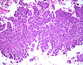
13756570 - Endometrioid carcinoma of the uterus, light micrograph
13619778 - Human vagina, light micrograph
13619740 - Human umbilical cord, light micrograph
13619712 - Rabbit corpus albicans, light micrograph
13619696 - Human cervix, light micrograph

13525015 - Childbirth, 19th century illustration
13524826 - Childbirth, 19th century illustration
13473113 - Oocyte, TEM

13473108 - Endoplasmic reticulum, TEM

13453162 - Female genitals
13452726 - Tubal ligation clips, X-ray
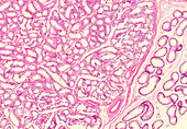
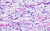
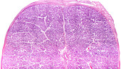
13416543 - Testicular cross section, light micrograph
13416541 - Ovarian section and corpus luteum, light micrograph

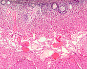
13416522 - Epididymal tuberculosis, light micrograph
13387038 - Cervical cancer, MRI scan
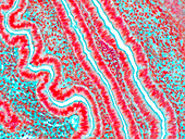
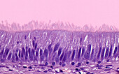
13356092 - Fallopian tube, TEM
13756594 - Serous carcinoma, light micrograph
13756590 - Carcinosarcoma, light micrograph

13619782 - Rabbit corpus luteum, light micrograph
13619775 - Human vagina, light micrograph
13619742 - Human umbilical cord, light micrograph
13619724 - Monkey oviduct, light micrograph
13525337 - Chicken embryo development, 19th century illustration

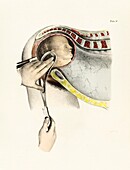
13525051 - Extracting retained placenta, 19th century illustration
13525048 - Breech delivery, 19th century illustration
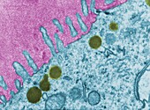
13501998 - Chlamydia inside epithelial cells of the oviduct, TEM

13473112 - Oocyte, TEM
13377735 - Endodermal sinus tumour, light micrograph
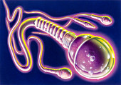
13376779 - Sperm cells, illustration
13296040 - Choriocarcinoma, light micrograph
13765240 - Uterus during menstruation, SEM
13756592 - Serous carcinoma, light micrograph
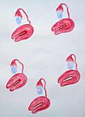
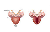
13685529 - Healthy and unhealthy prostate glands, illustration
13619716 - Monkey oviduct, light micrograph
13619707 - Human placenta, light micrograph
13476800 - Penile cancer, light micrograph
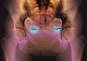
13452657 - Bicornuate uterus, X-ray
13218553 - Cancer of the womb, light micrograph
13619785 - Human Bartholin's gland, light micrograph
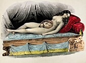
13525363 - Pregnant woman, 19th century illustration
13502257 - Chlamydia on surface of oviduct, TEM
13501995 - Chlamydia inside epithelial cells of the oviduct, TEM
13453114 - Lichen planus
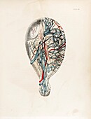
13443203 - Testicles and glass making instrument, illustration
13427199 - Penile skin cancer, light micrograph
13377741 - Chronic endometritis, light micrograph
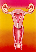
13376805 - Female reproductive system, illustration
nächste Seite
Fortpflanzungssystem Bilder ❘ Science Photo Library