Normal coronal and cross-sections of brain, illustration
Bildnummer 14167136
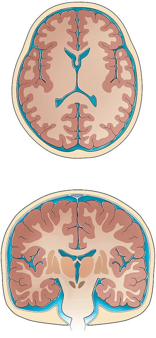
| Normal coronal and cross-sections of brain, illustration. Slices through the normal brain and skull showing ventricles, white matter, grey matter, basal ganglia, thalamus sulci and gyri, cerebrospinal fluid and skull. | |
| Lizenzart: | Lizenzpflichtig |
| Credit: | Science Photo Library / Sue Seif |
| Bildgröße: | 2841 px × 6150 px |
| Modell-Rechte: | nicht erforderlich |
| Eigentums-Rechte: | nicht erforderlich |
| Restrictions: | - |
Preise für dieses Bild ab 15 €
Universitäten & Organisationen
(Informationsmaterial Digital, Informationsmaterial Print, Lehrmaterial Digital etc.)
ab 15 €
Redaktionell
(Bücher, Bücher: Sach- und Fachliteratur, Digitale Medien (redaktionell) etc.)
ab 30 €
Werbung
(Anzeigen, Aussenwerbung, Digitale Medien, Fernsehwerbung, Karten, Werbemittel, Zeitschriften etc.)
ab 55 €
Handelsprodukte
(bedruckte Textilie, Kalender, Postkarte, Grußkarte, Verpackung etc.)
ab 75 €
Pauschalpreise
Rechtepakete für die unbeschränkte Bildnutzung in Print oder Online
ab 495 €