Cervical cancer cells dividing, SEM
Bildnummer 13954668
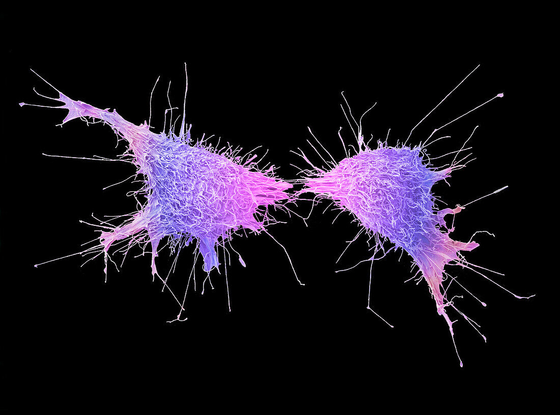
| Cervical cancer cells dividing, coloured scanning electron micrograph (SEM). The cervix is the lower part of the womb, also called the neck of the womb and comprises part of the womanâ??s reproductive system. Cervical cancer is more common in younger women and one of the main causes is a persistent infection of certain types of the human papilloma virus (HPV). In this image the cells are undergoing cytokinesis, which is the physical process of cell division that divides the parental cell into two daughter cells. Cancer cells often divide and multiply uncontrollably which can lead to the formation of tumours. At the end of cytokinesis, the two daughter cells remain connected by the midbody for a short time. The midbody is organised by a set of microtubules and its main function is to localise the site of natural detachment (abscission) between the two daughter cells. Magnification: x1600 when printed at 10cm wide | |
| Lizenzart: | Lizenzpflichtig |
| Credit: | Science Photo Library / ANNE E. WESTON |
| Bildgröße: | 3855 px × 2859 px |
| Modell-Rechte: | nicht erforderlich |
| Eigentums-Rechte: | nicht erforderlich |
| Restrictions: | - |
Preise für dieses Bild ab 15 €
Universitäten & Organisationen
(Informationsmaterial Digital, Informationsmaterial Print, Lehrmaterial Digital etc.)
ab 15 €
Redaktionell
(Bücher, Bücher: Sach- und Fachliteratur, Digitale Medien (redaktionell) etc.)
ab 30 €
Werbung
(Anzeigen, Aussenwerbung, Digitale Medien, Fernsehwerbung, Karten, Werbemittel, Zeitschriften etc.)
ab 55 €
Handelsprodukte
(bedruckte Textilie, Kalender, Postkarte, Grußkarte, Verpackung etc.)
ab 75 €
Pauschalpreise
Rechtepakete für die unbeschränkte Bildnutzung in Print oder Online
ab 495 €
Keywords
- Aktin,
- farbig,
- Gebärmutter,
- Gebärmutterhals,
- Gebärmutterhals-,
- gefärbt,
- Gesundheitswesen,
- HPV,
- Krankheit,
- Krebs,
- krebsartig,
- maligne,
- Medizin,
- medizinisch,
- mikroskopisch,
- Mikrotubuli,
- Papillom,
- Rasterelektronenmikroskopie,
- rasterelektronenmikroskopische Aufnahme,
- REM,
- schwarzer Hintergrund,
- Zelle,
- Zellen,
- Zytokinese,
- Zytologie