Bilder
Videos
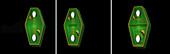
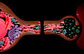
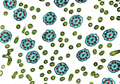
13951607 - Cytokinesis, illustration
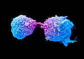
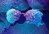
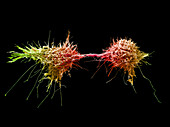
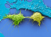
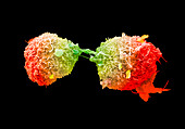
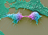
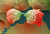
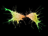
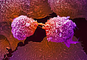
13742779 - Lung cancer cells dividing, SEM
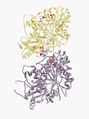
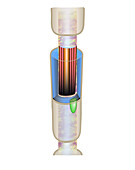
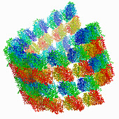
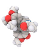
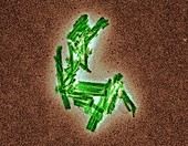
13406144 - Colchicine drug complexed with tubulin, molecular model
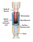
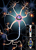
13950788 - Nerve cell axon anatomy, illustration
13755943 - Centriole, TEM
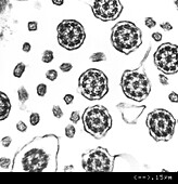
13755961 - Cilia, TEM
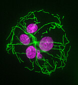
13632915 - Chromosomes during mitosis, illustration
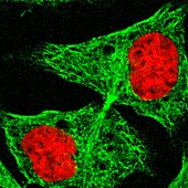
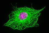
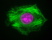
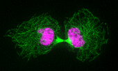
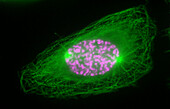
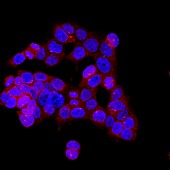
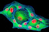
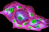
13243195 - Cancer cell showing tubulin, light micrograph
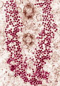
13755897 - Interphase cell treated with Colchicine, light micrograph
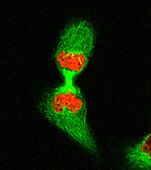
13755884 - Mitosis, light micrograph
13755802 - Ciliary doublet microtubule, illustration
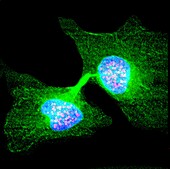
13672550 - Human cell late in cytokinesis, light micrograph

13632907 - Sperm cell, illustration
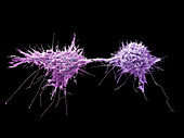
13742776 - Lung cancer cells dividing, SEM
13950789 - Nerve cell axon anatomy, illustration

13755938 - Cilia, TEM
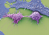
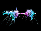
13686080 - Cervical cancer cells dividing, SEM
13672536 - Human cell early in cytokinesis, light micrograph
13672544 - Human cell in telophase, light micrograph

13954668 - Cervical cancer cells dividing, SEM
13755887 - Mitosis, light micrograph
13686084 - Cervical cancer cells dividing, SEM
13672554 - Human cell in telophase, light micrograph
13686083 - Cervical cancer cells dividing, SEM
13672557 - Two human cells in interphase, light micrograph
13755895 - Interphase cell treated with Taxol, light micrograph
13755889 - Mitosis, light micrograph
13742780 - Lung cancer cells dividing, SEM
13619616 - Nasal epithelial cells, TEM
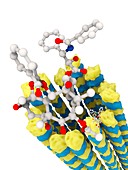
13838534 - Microtubule with tubulin oligomers, molecular model
13838533 - Microtubule with tubulin oligomers, molecular model
13686081 - Cervical cancer cells dividing, SEM
13950792 - Nerves cell and synapse structure, illustration
13755896 - Interphase cell treated with Colchicine, light micrograph
13755891 - Cytokinesis, light micrograph
13742777 - Lung cancer cells dividing, SEM
13674191 - Nassula sp. protozoa, light micrograph
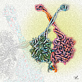
13954671 - Dynein-1 motor domain, illustration
13755886 - Mitosis, light micrograph

13405953 - Microtubule, computer model
13954667 - Cervical cancer cells dividing, SEM
13755890 - Mitosis, light micrograph
13686082 - Cervical cancer cells dividing, SEM
13356883 - Mitotic cell division, TEM
14077782 - Microtubules, TEM

13356877 - Platelet, TEM
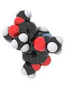
12967539 - Taxol molecule,illustration
13686085 - Cervical cancer cells dividing, SEM
13632837 - Chromosomes during mitosis, illustration
12967540 - Taxol and microtubule molecules,illustration
13406119 - Colchicine gout drug, molecular model
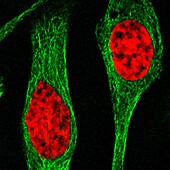
13243200 - Cancer cells cytoskeleton and nuclei, light micrograph
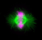
12968925 - Mitosis, confocal light micrograph
13387624 - Early clot formation, TEM
13295851 - Mitosis, illustration
13244416 - Intracellular transport of nanoparticles, illustration
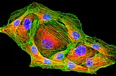
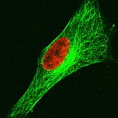
13435128 - HeLa cell, fluorescent light micrograph
13387574 - Early clot formation, TEM

13295853 - Mitosis, illustration
13244414 - Intracellular transport of nanoparticles, illustration
12967541 - Taxol and microtubule molecules,illustration
13954666 - Cervical cancer cells dividing, SEM
13755885 - Mitosis, light micrograph
13387560 - Early clot formation, TEM
12967535 - Taxol and microtubule molecules,illustration
13632855 - Microtrabecular network, illustration

13378272 - Tau protein clusters, TEM

11691451 - Plant cell mitosis,light micrograph
14077783 - Microtubules, TEM
13954669 - Cervical cancer cells dividing, SEM

13755888 - Mitosis, light micrograph
13686087 - Cervical cancer cells dividing, SEM
13672538 - Human cell in interphase, light micrograph
13632806 - Nerve cell axon anatomy, illustation
13378282 - Tau protein clusters, TEM
13243215 - Osteosarcoma cytoskeleton and nuclei, light micrograph

12968929 - Aberrant mitosis, confocal light micrograph
13406118 - Colchicine gout drug, molecular model
13356878 - Mitotic cell division, TEM
12950628 - Human cell, illustration
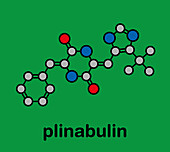
12917407 - Plinabulin cancer drug molecule
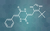
12917406 - Plinabulin cancer drug molecule
13755947 - Cilia and microvilli, TEM
13742778 - Lung cancer cells dividing, SEM
13686086 - Cervical cancer cells dividing, SEM
13243214 - Osteosarcoma Cells cytoskeleton and nuclei, light micrograph
12961095 - Sperm tails,TEM
12950454 - Microtubule construction, illustration

12916946 - Docetaxel chemotherapy drug molecule

13295849 - Mitosis, illustration
12362491 - Paramecium multimicronucleatum, TEM
13686363 - Respiratory epithelium, TEM
13405959 - Microtubule, computer model
12968924 - Mitosis, confocal light micrograph
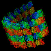
13406146 - Colchicine gout drug complexed with tubulin, molecular model
13295850 - Mitosis, illustration
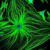
12968931 - Astrocyte brain cells, confocal light micrograph
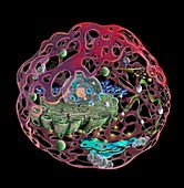
12950629 - Human cell, illustration
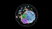
13959108 - Human cell, illustration
nächste Seite
Mikrotubuli Bilder ❘ Science Photo Library