Retina damage from diabetes, illustration
Bildnummer 13635996
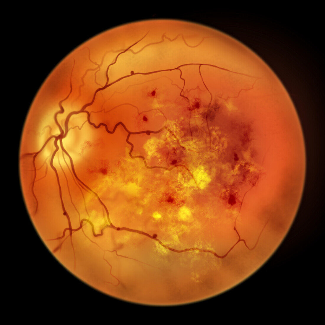
| Retina damage from diabetes. Computer illustration showing an ophthalmoscope view of hard exudates (accumulation of fatty substances leaked from blocked capillaries seen as small yellow dots), cotton wool spots (accumulation of axoplasmic material within the nerve fibre layer seen as fluffy yellow patches), haemorrhages and microaneurysms (red spots), macula oedema. | |
| Lizenzart: | Lizenzfrei |
| Credit: | Science Photo Library / Kon, Kateryna |
| Modell-Rechte: | nicht erforderlich |
| Eigentums-Rechte: | nicht erforderlich |
| Restrictions: | - |
Preise für dieses Bild ab 29 €
Für digitale Nutzung (72 dpi)
ab 29 €
Für Druckauflösung (300 dpi)
ab 300 €
Keywords
- abnormal,
- Auge,
- Augen-,
- Augenheilkunde,
- Bluten,
- Blutgefäße,
- Close-up,
- Detail,
- Diabetes,
- Diabetiker,
- diabetische Retinopathie,
- Erwachsene,
- Fundoskopie,
- geduldig,
- Illustration,
- kapillar,
- Kondition,
- Krankheit,
- Kreislauf,
- Kunstwerk,
- linkes Auge,
- Medizin,
- medizinisch,
- menschlicher Körper,
- Netzhaut-,
- Niemand,
- Retina,
- schwarzer Hintergrund,
- Störung,
- ungesund,
- vaskulär