Bilder
Videos
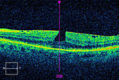
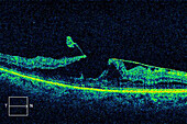
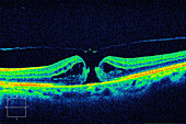
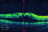
13687022 - Macular pucker with lamellar hole, OCT scan
13687021 - Macular pucker with lamellar hole, OCT scan
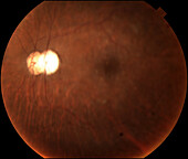
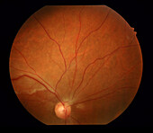
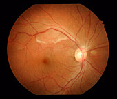
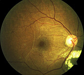
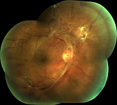
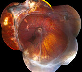
13613554 - Intraocular lens subluxation, fundoscopy
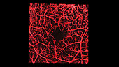
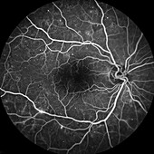
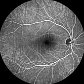
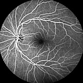
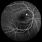
13951663 - Retinal blood vessels, OCT angiography scan
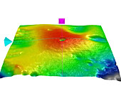
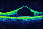
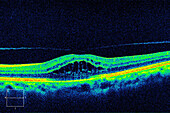
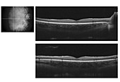
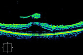
13733185 - Diabetic macular oedema, OCT macular cube scan
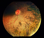
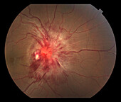
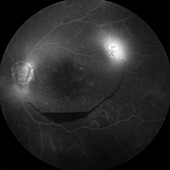
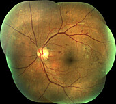
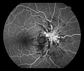
13613586 - Retinal vasculitis, fundoscopy
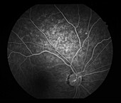
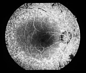
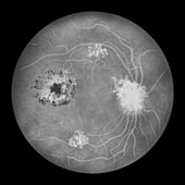
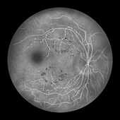
13613568 - Retina damage from diabetes, angiogram

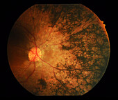
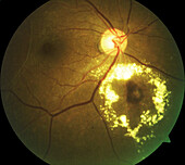
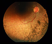
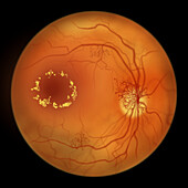
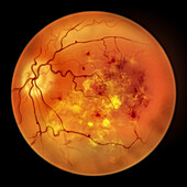
13613650 - Retina damage from diabetes, fundoscopy
13613640 - Retina damage from diabetes, angiogram
13613577 - Retinal vasculitis, fundoscopy
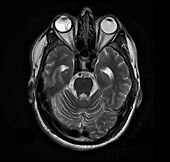
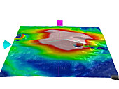
13599593 - Retinal detachment, MRI scan
13733188 - Macular pucker with lamellar hole, OCT macular cube scan
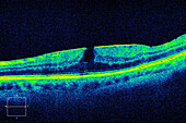
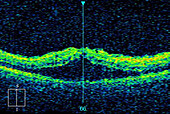
13613558 - Vitreomacular traction, OCT scan

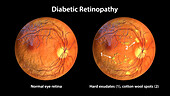
13736716 - Healthy eye and diabetic retinopathy, illustration
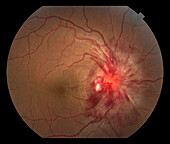
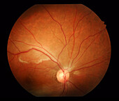
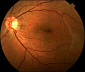
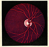
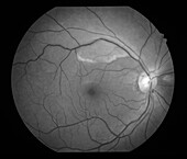
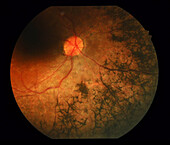
13613564 - Papilloedema, fundoscopy
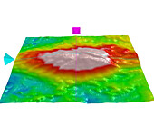
13733186 - Diabetic macular oedema, OCT macular cube scan
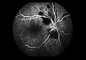
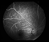
13732016 - Anterior ischemic optic neuropathy, angiogram
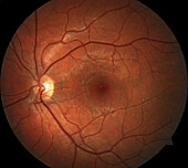
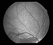
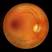
13613550 - Healthy eye, fundoscopy
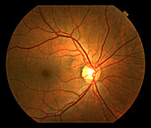
13613548 - Healthy eye, fundoscopy
13732018 - Temporal epiretinal membranes, fundoscopy
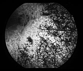
13613634 - Pigmentary retinopathy, fundoscopy
13613632 - Pigmentary retinopathy, fundoscopy
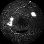
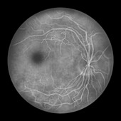
13613546 - Healthy eye, angiogram
13687019 - Diabetic macular oedema, OCT scan
13687015 - Diabetic macular oedema, OCT scan
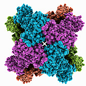
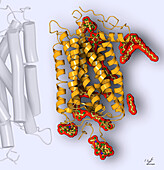
13686331 - Human retinal IMPDH1 variant, molecular model

13613545 - Healthy eye, angiogram

13687016 - Diabetic macular oedema, OCT scan
13613581 - Retinal vasculitis, fundoscopy

13733184 - Diabetic macular oedema, OCT macular cube scan

13736715 - Healthy eye and diabetic retinopathy, illustration
13687020 - Lamellar macular hole, OCT scan
13613547 - Healthy eye, angiogram
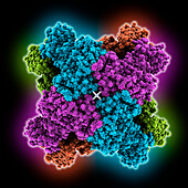
13686330 - Human retinal IMPDH1 variant, molecular model
13732017 - Central serous retinopathy, angiogram

13613584 - Retinal vasculitis, angiogram
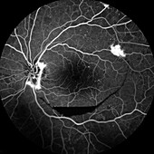
13613570 - Retina damage from diabetes, angiogram
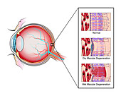
13736723 - Macular degeneration, illustration

13736722 - Macular degeneration, illustration

13673066 - Structure of the eye, illustration

13613647 - Retina damage from diabetes, angiogram
13613638 - Pigmentary retinopathy, angiogram
13613582 - Retinal vasculitis, fundoscopy
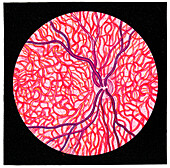
13736133 - Normal fundus, illustration
13613654 - Vitreomacular traction, fundoscopy
13613565 - Papilloedema, fundoscopy
13357198 - Retina plexiform layer, TEM
13613572 - Retina damage from diabetes, angiogram
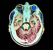
13599595 - Retinal detachment, MRI scan

13376764 - Eye anatomy, illustration
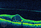
13687018 - Diabetic macular oedema, OCT scan
13613653 - Vitreomacular traction, fundoscopy
13613648 - Retina damage from diabetes, fundoscopy
13742358 - Rhodopsin, illustration
13687017 - Cystoid macular oedema, OCT scan
13687014 - Diabetic macular oedema, OCT scan
13636025 - Retina damage from diabetes, illustration
13613636 - Pigmentary retinopathy, fundoscopy
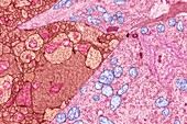
13357186 - Retina plexiform layer, TEM

13224879 - Facial recognition, illustration
13613567 - Papilloedema, angiogram
13613649 - Retina damage from diabetes, fundoscopy
13613583 - Retinal vasculitis, angiogram
13636058 - Retina damage from diabetes, illustration
13613588 - Retinal vasculitis, fundoscopy
12646750 - Spectral Domain optical coherence tomography of human fovea
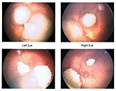
13613664 - Benign eye tumour, fundoscopy
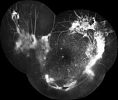
13613651 - Retina damage from diabetes, angiogram
13613635 - Pigmentary retinopathy, fundoscopy
13613585 - Retinal vasculitis, angiogram
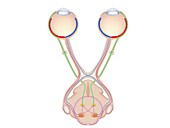
12971340 - Brain's visual pathways, illustration
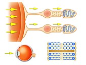
12971338 - Biochemistry of the retina, illustration
12647748 - Retina, LM

13838915 - Retinal cyclic nucleotide-gated channel, illustration

13613667 - Benign eye tumour, fundoscopy
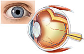
12648556 - Eye Anatomy, illustration
13733187 - Diabetic macular oedema, OCT macular cube scan
13733176 - Fundus in ocular albinism, illustration
13613637 - Pigmentary retinopathy, angiogram
13613555 - Vitreomacular traction, OCT scan
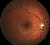
13613549 - Healthy eye, fundoscopy

13599594 - Retinal detachment, MRI scan

13416710 - Human retinal organoid, fluorescent micrograph
12649522 - Retinoblastoma Scan Before and After Chemotherapy
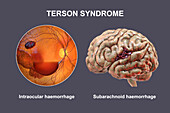
14079774 - Terson syndrome, illustration
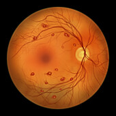
14079588 - Roth spots in the retina, illustration
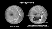
14079557 - Intraocular haemorrhage in Terson syndrome, illustration
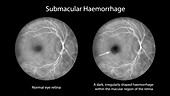
14079545 - Submacular haemorrhage on the retina, illustration
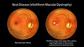
13956055 - Best disease, illustration
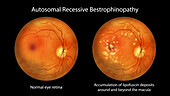
13956052 - Autosomal recessive bestrophinopathy, illustration
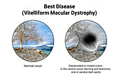
13956030 - Normal eye and an eye with Best disease, illustration
13636093 - Retina damage from diabetes, illustration

13636065 - Retina damage from diabetes, illustration
13636059 - Retina damage from diabetes, illustration
13636016 - Retina damage from diabetes, illustration
13636010 - Retina damage from diabetes, illustration
13635996 - Retina damage from diabetes, illustration

13635239 - Microaneurysms, illustration
13635238 - Microaneurysms, illustration
nächste Seite
Netzhaut- Bilder ❘ Science Photo Library