Sperm, SEM-TEM comparison
Bildnummer 12948529
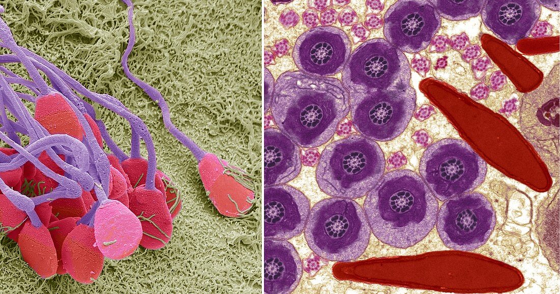
| Sperm cells. Comparison between a scanning electron micrograph (SEM, left) and transmission electron micrograph (TEM, right) of sperm cells. In the TEM image the heads of the sperm, which contain the genetic material, are bottom and right (red). The central part of the cells contains mitochondria and provides the energy for the cell, (round, larger). The ends of the tails are the smallest round structures. The tail's motility is aided by the arrangement of microtubules (part of the cell cytoskeleton) within the tail. The microtubules are arranged in a central axoneme, with a central pair of microtubules surrounded by nine other pairs in a ring. Magnification: SEM x3000, TEM x10, 000 when printed 10 centimetres high. For a series of comparisons between SEMs and TEMs see images C047/7006 to C047/7034. | |
| Lizenzart: | Lizenzpflichtig |
| Credit: | Science Photo Library / Gschmeissner, Steve |
| Bildgröße: | 8157 px × 4285 px |
| Modell-Rechte: | nicht erforderlich |
| Eigentums-Rechte: | nicht erforderlich |
| Restrictions: | - |
Preise für dieses Bild ab 15 €
Universitäten & Organisationen
(Informationsmaterial Digital, Informationsmaterial Print, Lehrmaterial Digital etc.)
ab 15 €
Redaktionell
(Bücher, Bücher: Sach- und Fachliteratur, Digitale Medien (redaktionell) etc.)
ab 30 €
Werbung
(Anzeigen, Aussenwerbung, Digitale Medien, Fernsehwerbung, Karten, Werbemittel, Zeitschriften etc.)
ab 55 €
Handelsprodukte
(bedruckte Textilie, Kalender, Postkarte, Grußkarte, Verpackung etc.)
ab 75 €
Pauschalpreise
Rechtepakete für die unbeschränkte Bildnutzung in Print oder Online
ab 495 €
Keywords
- Anatomie,
- anatomisch,
- Axonem,
- Entwicklung,
- farbig,
- Fauna,
- gefärbt,
- gesund,
- Keimzelle,
- männliche Fortpflanzungszelle,
- Mikrotubuli,
- Mitochondrien,
- normal,
- Rasterelektronenmikroskop,
- rasterelektronenmikroskopische Aufnahme,
- Reihenfolge,
- REM,
- Reproduktion,
- Serie,
- Spermatozoen,
- Spermatozoon,
- tem,
- Tier,
- Tierwelt,
- Transmissionselektronenmikroskop,
- transmissionselektronenmikroskopische Aufnahme,
- Vergleich,
- vergleichen,
- verglichen,
- Zellen,
- Zytoskelett