Mitosis cell division
Bildnummer 12071198
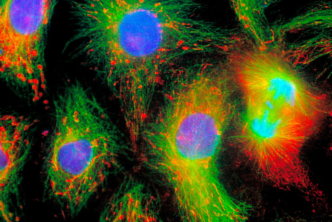
| Mitosis. Immunofluorescent light micrograph of kidney cells from a rat kangaroo (Potorous tridactylus). The cell at far right is seen during the anaphase stage of mitotic cell division. The two chromatids that form each chromosome are being pulled to opposite ends of the cell along spindle fibres. The other cells are seen during interphase, the stage between mitotic cell divisions. The chromosomes are located in the nucleus (purple). They are surrounded by actin microfilaments (red) and tubulin microtubules (green) of the cytoskeleton that maintains the structure and organisation of the cell. Magnification: x2700 when printed 10cm wide. | |
| Lizenzart: | Lizenzpflichtig |
| Credit: | Science Photo Library / Waters, Jennifer |
| Bildgröße: | 6040 px × 4040 px |
| Modell-Rechte: | nicht erforderlich |
| Eigentums-Rechte: | nicht erforderlich |
| Restrictions: |
|
Preise für dieses Bild ab 15 €
Universitäten & Organisationen
(Informationsmaterial Digital, Informationsmaterial Print, Lehrmaterial Digital etc.)
ab 15 €
Redaktionell
(Bücher, Bücher: Sach- und Fachliteratur, Digitale Medien (redaktionell) etc.)
ab 30 €
Werbung
(Anzeigen, Aussenwerbung, Digitale Medien, Fernsehwerbung, Karten, Werbemittel, Zeitschriften etc.)
ab 55 €
Handelsprodukte
(bedruckte Textilie, Kalender, Postkarte, Grußkarte, Verpackung etc.)
ab 75 €
Pauschalpreise
Rechtepakete für die unbeschränkte Bildnutzung in Print oder Online
ab 495 €