Mitosis cell division
Bildnummer 11874822
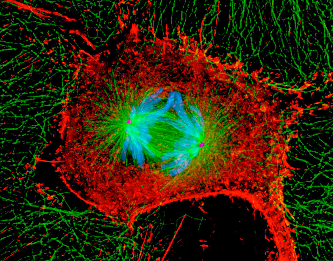
| Mitosis (image 4 of 6). Digital three-dimensional immunofluorescent light micrograph of a section through a mammalian cell during the anaphase stage of mitotic cell division. The two chromatids that form each chromosome (blue) are being pulled to opposite ends of the cell. The actin microfilam- ents (red) and tubulin microtubules (green) of the cytoskeleton maintain the cell's structure. The two centrosomes (pink dots,centre left & right) organize the microtubules that move the chromosom- es around the cell. Mitosis produces two identical daughter cells. This is a rat kangaroo kidney epi- thelial cell. Magnification: x500 at 6x7cm size. For a sequence of mitosis see images P673/052-057 | |
| Lizenzart: | Lizenzpflichtig |
| Credit: | Science Photo Library / Khodjakov, Dr. Alexey |
| Bildgröße: | 3500 px × 2742 px |
| Modell-Rechte: | nicht erforderlich |
| Eigentums-Rechte: | nicht erforderlich |
| Restrictions: | - |
Preise für dieses Bild ab 15 €
Universitäten & Organisationen
(Informationsmaterial Digital, Informationsmaterial Print, Lehrmaterial Digital etc.)
ab 15 €
Redaktionell
(Bücher, Bücher: Sach- und Fachliteratur, Digitale Medien (redaktionell) etc.)
ab 30 €
Werbung
(Anzeigen, Aussenwerbung, Digitale Medien, Fernsehwerbung, Karten, Werbemittel, Zeitschriften etc.)
ab 55 €
Handelsprodukte
(bedruckte Textilie, Kalender, Postkarte, Grußkarte, Verpackung etc.)
ab 75 €
Pauschalpreise
Rechtepakete für die unbeschränkte Bildnutzung in Print oder Online
ab 495 €