Mitosis
Bildnummer 11874741
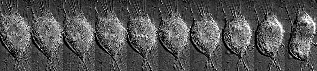
| Cell division. Time-exposure light micrograph showing the different stages in mitosis (nuclear division). At left the cell is in interphase. The nucleus (round,centre) is clearly visible. As the cell moves into prophase,the nuclear envelope dissolves and the chromosomes (white) condense. By the fifth frame,the cell is in metaphase,with the chromosomes aligned along the centre of the cell. The chromosomes start to move to the opposite poles,guided by microtubules,during anaphase. In the last frame the separated chromosomes have moved to opposite ends of the cell. The cell is undergoing cytokinesis (cell division) to form two new cells | |
| Lizenzart: | Lizenzpflichtig |
| Credit: | Science Photo Library / DR PAUL ANDREWS, UNIVERSITY OF DUNDEE |
| Bildgröße: | 6210 px × 1406 px |
| Modell-Rechte: | nicht erforderlich |
| Eigentums-Rechte: | nicht erforderlich |
| Restrictions: | - |
Preise für dieses Bild ab 15 €
Universitäten & Organisationen
(Informationsmaterial Digital, Informationsmaterial Print, Lehrmaterial Digital etc.)
ab 15 €
Redaktionell
(Bücher, Bücher: Sach- und Fachliteratur, Digitale Medien (redaktionell) etc.)
ab 30 €
Werbung
(Anzeigen, Aussenwerbung, Digitale Medien, Fernsehwerbung, Karten, Werbemittel, Zeitschriften etc.)
ab 55 €
Handelsprodukte
(bedruckte Textilie, Kalender, Postkarte, Grußkarte, Verpackung etc.)
ab 75 €
Pauschalpreise
Rechtepakete für die unbeschränkte Bildnutzung in Print oder Online
ab 495 €
Keywords
- Atomkern,
- Biologie,
- biologisch,
- Chromosom,
- Chromosomen,
- Desoxiribonukleinsäure,
- DNA,
- Einfarbig,
- Einteilung,
- Genetik,
- Interphase,
- Kerne,
- Krebszellen,
- Langzeitbelichtung,
- mehrere,
- Mikrotubuli,
- Mitose,
- nuklear,
- Panorama-,
- Prometaphase,
- Reihenfolge,
- Schwarz und weiß,
- Separieren,
- Teilen,
- Tochter,
- viele,
- Zelle,
- zellular,
- Zytokinese,
- Zytologie,
- Zytologisch