Dental X-ray
Bildnummer 11858975
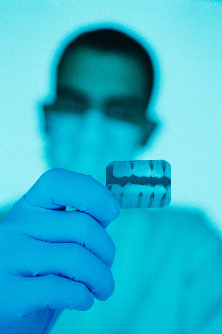
| Dental X-ray. Dentist holding a dental X-ray. It shows teeth (blue) from both the upper and lower jaw. The X-ray film is mounted in a device that is clenched between the teeth,positioning the X-ray film parallel to the teeth. An X-ray machine sends X-rays through the teeth to hit the film. This is known as exposing the film,and this image of the teeth is created on the film as the teeth block varying amounts of X-rays. This allows a dentist to assess the internal integrity of the teeth,and to check for any signs of tooth decay. The bright white areas in the teeth may be fillings or crowns that repaired previous areas of tooth decay. The black spot could be a tooth cavity | |
| Lizenzart: | Lizenzpflichtig |
| Credit: | Science Photo Library / Lawry, Lawrence |
| Bildgröße: | 3537 px × 5313 px |
| Modell-Rechte: | nicht erforderlich |
| Eigentums-Rechte: | nicht erforderlich |
| Restrictions: | - |
Preise für dieses Bild ab 15 €
Universitäten & Organisationen
(Informationsmaterial Digital, Informationsmaterial Print, Lehrmaterial Digital etc.)
ab 15 €
Redaktionell
(Bücher, Bücher: Sach- und Fachliteratur, Digitale Medien (redaktionell) etc.)
ab 30 €
Werbung
(Anzeigen, Aussenwerbung, Digitale Medien, Fernsehwerbung, Karten, Werbemittel, Zeitschriften etc.)
ab 55 €
Handelsprodukte
(bedruckte Textilie, Kalender, Postkarte, Grußkarte, Verpackung etc.)
ab 75 €
Pauschalpreise
Rechtepakete für die unbeschränkte Bildnutzung in Print oder Online
ab 495 €
Keywords
- Arbeit,
- Arbeiten,
- Arbeiter,
- Ausrüstung,
- Blau,
- dental,
- Diagnose,
- Erwachsene,
- Film,
- Füllung,
- Gesundheitswesen,
- Hand,
- Hohlraum,
- Karies,
- Krankheit,
- Loch,
- Mann,
- Männlich,
- Maske,
- maskiert,
- Medizin,
- medizinisch,
- Mensch,
- Mundhygiene,
- Person,
- Röntgen,
- Röntgenbild,
- Technologie,
- Zahn,
- Zahnarzt,
- Zähne,
- Zahnheilkunde,
- zerfallen