Cancer cell division
Bildnummer 11837465
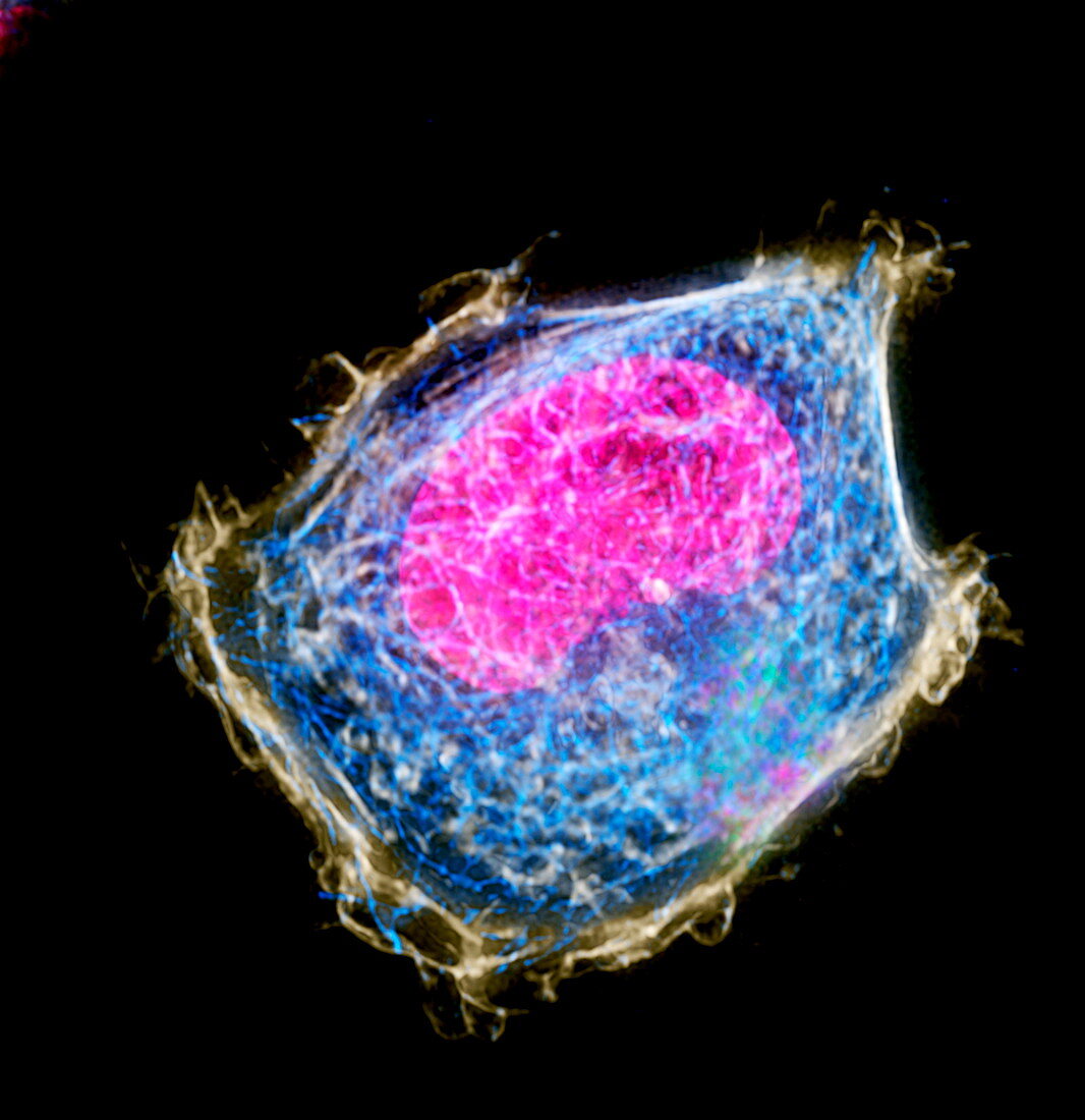
| Cancer cell division. Immunofluorescent light micrograph of a cancer cell during the interphase stage of cell division (mitosis). Interphase is the resting stage of mitosis,when the cell's deoxyribonucleic acid (DNA,pink) is replicated. Also seen is the actin cytoskeleton (white) and the microtubules (blue). During interphase,proteins are also synthesised and the cell increases in size. During mitosis a cell produces two genetically- identical daughter cells. Immunofluorescence uses antibodies to attach fluorescent dyes to specific cell tissues | |
| Lizenzart: | Lizenzpflichtig |
| Credit: | Science Photo Library / DR PAUL ANDREWS, UNIVERSITY OF DUNDEE |
| Bildgröße: | 2635 px × 2718 px |
| Modell-Rechte: | nicht erforderlich |
| Eigentums-Rechte: | nicht erforderlich |
| Restrictions: | - |
Preise für dieses Bild ab 15 €
Universitäten & Organisationen
(Informationsmaterial Digital, Informationsmaterial Print, Lehrmaterial Digital etc.)
ab 15 €
Redaktionell
(Bücher, Bücher: Sach- und Fachliteratur, Digitale Medien (redaktionell) etc.)
ab 30 €
Werbung
(Anzeigen, Aussenwerbung, Digitale Medien, Fernsehwerbung, Karten, Werbemittel, Zeitschriften etc.)
ab 55 €
Handelsprodukte
(bedruckte Textilie, Kalender, Postkarte, Grußkarte, Verpackung etc.)
ab 75 €
Pauschalpreise
Rechtepakete für die unbeschränkte Bildnutzung in Print oder Online
ab 495 €
Keywords
- abnormal,
- Aktin-Zytoskelett,
- Ausruhen,
- Biologie,
- biologisch,
- Bühne,
- Desoxiribonukleinsäure,
- DNA,
- einer,
- Genetik,
- Interphase,
- kopierend,
- Krebs,
- krebsartig,
- Lichtmikroskop,
- lichtmikroskopische Aufnahme,
- Mikrotubuli,
- Mitose,
- Onkologie,
- onkologisch,
- Phase,
- ruhen,
- Single,
- ungesund,
- Zelle,
- Zytologie,
- Zytologisch