Ultraviolet fluorescence LM of BHK Cells
Bildnummer 11821561
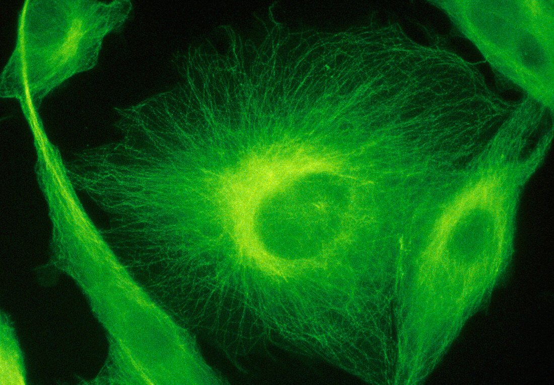
| Ultraviolet fluorescence light micrograph of BHK (baby hampster kidney) tissue culture cells labelled with anti-tubulin to show the micro- tubular structure. Microtubules,together with microfilaments (not seen here),form a three- dimensional array known as the cytoskeleton. This fibrous network,only recently observed using ultraviolet light & fluorescent stains,is still poorly understood. Microtubules,more rigid than microfilaments,are thought to act as direction markers in the cell. In this image the oval depression at the centre of each cell is the space occupied by the nucleus. The microtubules radiate from the nucleus to the periphery of the cell | |
| Lizenzart: | Lizenzpflichtig |
| Credit: | Science Photo Library / Murti, Dr. Gopal |
| Bildgröße: | 3729 px × 2594 px |
| Modell-Rechte: | nicht erforderlich |
| Eigentums-Rechte: | nicht erforderlich |
| Restrictions: | - |
Preise für dieses Bild ab 15 €
Universitäten & Organisationen
(Informationsmaterial Digital, Informationsmaterial Print, Lehrmaterial Digital etc.)
ab 15 €
Redaktionell
(Bücher, Bücher: Sach- und Fachliteratur, Digitale Medien (redaktionell) etc.)
ab 30 €
Werbung
(Anzeigen, Aussenwerbung, Digitale Medien, Fernsehwerbung, Karten, Werbemittel, Zeitschriften etc.)
ab 55 €
Handelsprodukte
(bedruckte Textilie, Kalender, Postkarte, Grußkarte, Verpackung etc.)
ab 75 €
Pauschalpreise
Rechtepakete für die unbeschränkte Bildnutzung in Print oder Online
ab 495 €