Mitosis,3D-structured micrograph
Bildnummer 11673097
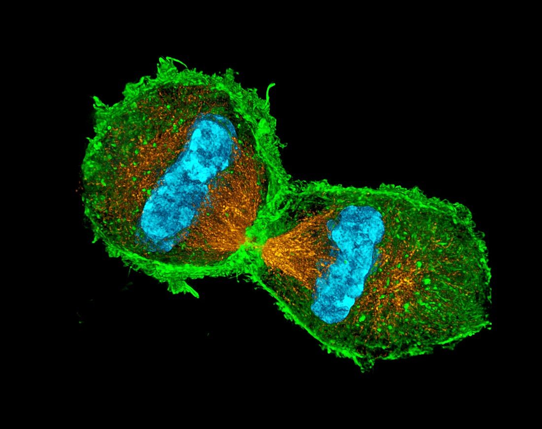
| Mitosis. 3D-structured illumination micrograph of the nucleus of a mouse fibroblast cell in the telophase,the last stage of mitosis (nuclear division). During mitosis two daughter nuclei are formed from one parent nucleus. The chromosomes (blue) have been split into their identical sister chromatids and moved to separate poles of the cell by microtubules (orange). This means that each daughter cell retains the parent cell's genetic information. A actin (green) ring is forming in preparation for cytokinesis (cell division) | |
| Lizenzart: | Lizenzpflichtig |
| Credit: | Science Photo Library / Schermelleh, Dr. Lothar |
| Bildgröße: | 3938 px × 3118 px |
| Modell-Rechte: | nicht erforderlich |
| Eigentums-Rechte: | nicht erforderlich |
| Restrictions: | - |
Preise für dieses Bild ab 15 €
Universitäten & Organisationen
(Informationsmaterial Digital, Informationsmaterial Print, Lehrmaterial Digital etc.)
ab 15 €
Redaktionell
(Bücher, Bücher: Sach- und Fachliteratur, Digitale Medien (redaktionell) etc.)
ab 30 €
Werbung
(Anzeigen, Aussenwerbung, Digitale Medien, Fernsehwerbung, Karten, Werbemittel, Zeitschriften etc.)
ab 55 €
Handelsprodukte
(bedruckte Textilie, Kalender, Postkarte, Grußkarte, Verpackung etc.)
ab 75 €
Pauschalpreise
Rechtepakete für die unbeschränkte Bildnutzung in Print oder Online
ab 495 €
Keywords
- Aktin,
- Biologie,
- biologisch,
- Bühne,
- Chromosom,
- Chromosomen,
- Dreidimensional,
- Fibroblasten,
- Fluoreszenz,
- fluoreszierend,
- Genetik,
- kopierend,
- Lichtmikroskop,
- lichtmikroskopische Aufnahme,
- Mikrofotografie,
- Mikrotubuli,
- Mitose,
- Ring,
- schwarzer Hintergrund,
- Spindel,
- Teilen,
- Zellbilogie,
- Zellkern,
- Zytologie,
- Zytologisch