Vaccinia virus infected cell
Bildnummer 11603489
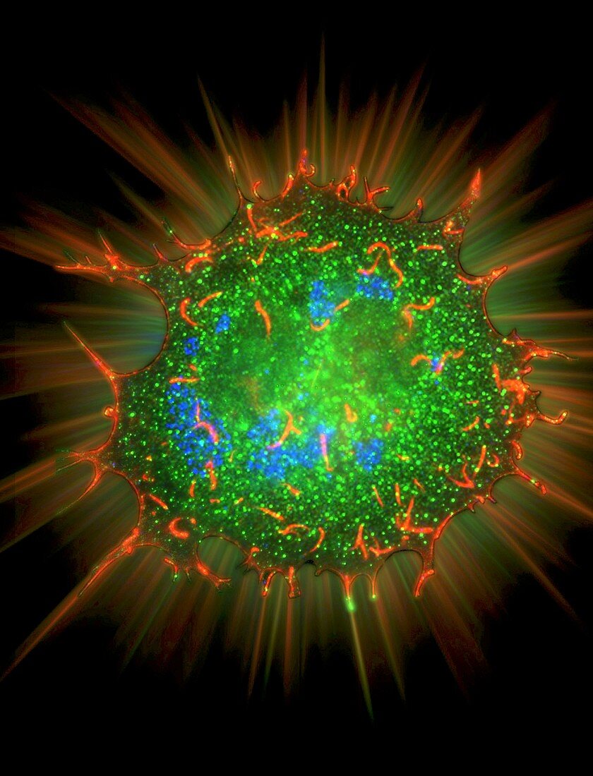
| Vaccinia virus infected cell. Immunofluorescence deconvolution micrograph of a cell infected with vaccinia virus particles. Viral DNA (deoxyribonucleic acid) is blue and its presence highlights areas of virus assembly within the cell. Actin protein filaments,which make up part of the cytoskeleton,are red. The cytoskeleton maintains the cell's shape and is involved in intracellular transport. The virus uses the actin to propel the newly formed particles out of the cell,forming the protrusions seen here. The tyrosine kinase c-Abl,which is involved in catalysing actin motility,is green | |
| Lizenzart: | Lizenzpflichtig |
| Credit: | Science Photo Library / Kalman, Dr. Dan / Vicari, Katie |
| Bildgröße: | 2598 px × 3404 px |
| Modell-Rechte: | nicht erforderlich |
| Eigentums-Rechte: | nicht erforderlich |
| Restrictions: | - |
Preise für dieses Bild ab 15 €
Universitäten & Organisationen
(Informationsmaterial Digital, Informationsmaterial Print, Lehrmaterial Digital etc.)
ab 15 €
Redaktionell
(Bücher, Bücher: Sach- und Fachliteratur, Digitale Medien (redaktionell) etc.)
ab 30 €
Werbung
(Anzeigen, Aussenwerbung, Digitale Medien, Fernsehwerbung, Karten, Werbemittel, Zeitschriften etc.)
ab 55 €
Handelsprodukte
(bedruckte Textilie, Kalender, Postkarte, Grußkarte, Verpackung etc.)
ab 75 €
Pauschalpreise
Rechtepakete für die unbeschränkte Bildnutzung in Print oder Online
ab 495 €
Keywords
- Aktin,
- Assembler-,
- Atomkern,
- befleckt,
- Biochemie,
- biochemisch,
- Biologie,
- biologisch,
- c-abl,
- Desoxiribonukleinsäure,
- DNA,
- Eiweiß,
- Entfaltung,
- Erreger,
- Farbstoff,
- Filament,
- Filamente,
- fluoreszierend,
- Infektion,
- infiziert,
- konfokale mikroskopische Aufnahme,
- kopierend,
- Kuhpockenvirus,
- Lichtmikroskop,
- lichtmikroskopische Aufnahme,
- Medizin,
- medizinisch,
- Mikrobiologie,
- mikrobiologisch,
- Mikrotubuli,
- Replikation,
- Reproduktion,
- schwarzer Hintergrund,
- Vaccinia,
- Verfärbung,
- viral,
- Virologie,
- Virus,
- Zelle,
- Zytoskelett