Retina in glaucoma,artwork
Bildnummer 11554973
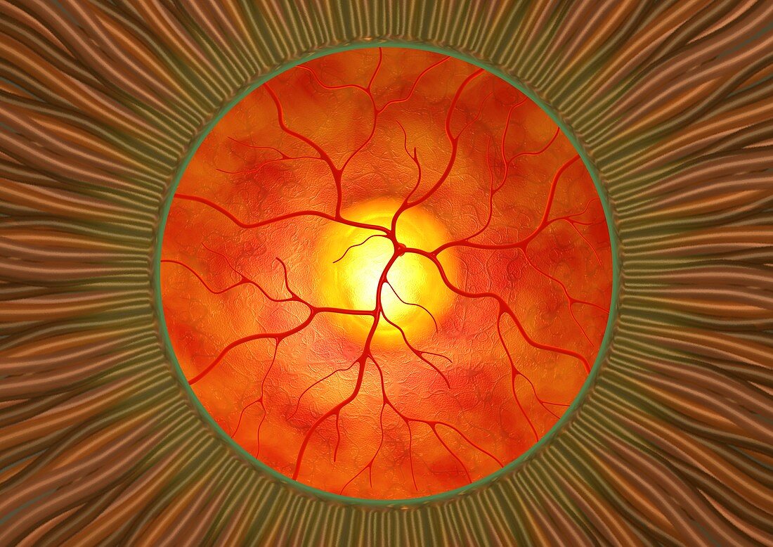
| Retina in glaucoma,computer artwork. This view is looking through the pupil (green ring) at the front of the eye,to the retina (orange) at the back of the eye. In glaucoma,the blood vessels (red) and the optic disc (yellow,head of the optic nerve) are compressed by increased fluid pressure inside the eye. The optic disc is raised with a central bulge or 'cup' (white). The cup-to-disc ratio is used to measure the progression of glaucoma. If left untreated glaucoma can cause impaired vision and eventually total blindness. The strands surrounding the pupil are part of the iris | |
| Lizenzart: | Lizenzpflichtig |
| Credit: | Science Photo Library / Animated Healthcare |
| Bildgröße: | 4984 px × 3524 px |
| Modell-Rechte: | nicht erforderlich |
| Eigentums-Rechte: | nicht erforderlich |
| Restrictions: | - |
Preise für dieses Bild ab 15 €
Universitäten & Organisationen
(Informationsmaterial Digital, Informationsmaterial Print, Lehrmaterial Digital etc.)
ab 15 €
Redaktionell
(Bücher, Bücher: Sach- und Fachliteratur, Digitale Medien (redaktionell) etc.)
ab 30 €
Werbung
(Anzeigen, Aussenwerbung, Digitale Medien, Fernsehwerbung, Karten, Werbemittel, Zeitschriften etc.)
ab 55 €
Handelsprodukte
(bedruckte Textilie, Kalender, Postkarte, Grußkarte, Verpackung etc.)
ab 75 €
Pauschalpreise
Rechtepakete für die unbeschränkte Bildnutzung in Print oder Online
ab 495 €