Blood and fibrin, light micrograph
Bildnummer 14113441
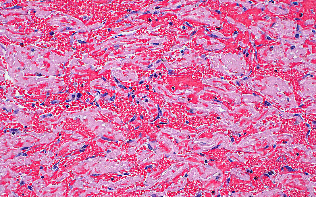
| Light micrograph of red blood cells and fibrin. The red blood cells (many small bright red dots) clump together with fibrin (light pink) in a blood clot. Scattered fibroblast nuclei (blue elongated dots) are also seen, indicating organisation or the cellular reaction to a clot that has been present in the body for some time. Haematoxylin and eosin stained tissue section. Magnification: 200x when printed at 10cm. | |
| Lizenzart: | Lizenzpflichtig |
| Credit: | Science Photo Library / ZIAD M. EL-ZAATARI |
| Bildgröße: | 4096 px × 2560 px |
| Modell-Rechte: | nicht erforderlich |
| Eigentums-Rechte: | nicht erforderlich |
| Restrictions: | - |
Preise für dieses Bild ab 15 €
Universitäten & Organisationen
(Informationsmaterial Digital, Informationsmaterial Print, Lehrmaterial Digital etc.)
ab 15 €
Redaktionell
(Bücher, Bücher: Sach- und Fachliteratur, Digitale Medien (redaktionell) etc.)
ab 30 €
Werbung
(Anzeigen, Aussenwerbung, Digitale Medien, Fernsehwerbung, Karten, Werbemittel, Zeitschriften etc.)
ab 55 €
Handelsprodukte
(bedruckte Textilie, Kalender, Postkarte, Grußkarte, Verpackung etc.)
ab 75 €
Pauschalpreise
Rechtepakete für die unbeschränkte Bildnutzung in Print oder Online
ab 495 €
Keywords
- abnormal,
- Anatomie,
- Blut,
- Blutgerinnsel,
- Gewebe,
- Histopathologie,
- histopathologisch,
- kardiovaskular,
- Kondition,
- Krankheit,
- Kreislauf,
- lichtmikroskopische Aufnahme,
- Medizin,
- medizinisch,
- menschlicher Körper,
- Mikroskopie,
- Niemand,
- Pathologie,
- pathologisch,
- rote Blutkörperchen,
- rutschen,
- Störung,
- Thrombus,
- ungesund,
- Zellen