Gallbladder cancer, light micrograph
Bildnummer 14113279
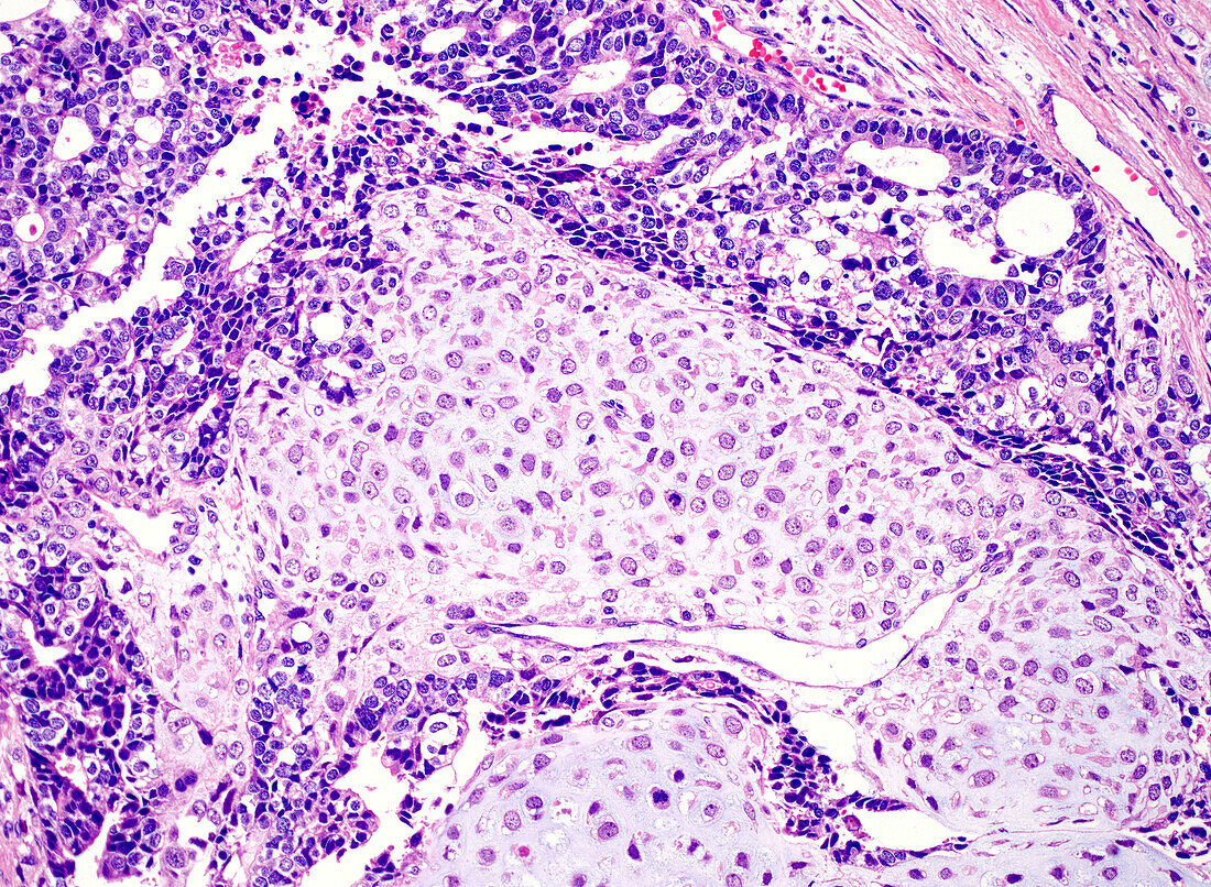
| Light micrograph of a haematoxylin and eosin-stained section from a gallbladder cancer. It is an example of a sarcomatoid carcinoma (also called carcinosarcoma) of the gallbladder. The carcinoma component consists of fused glandular structures (dark blue) which surround an area of cartilage (light blue). Cartilage represents heterologous differentiation in this case. | |
| Lizenzart: | Lizenzpflichtig |
| Credit: | Science Photo Library / WEBPATHOLOGY |
| Bildgröße: | 4096 px × 3000 px |
| Modell-Rechte: | nicht erforderlich |
| Eigentums-Rechte: | nicht erforderlich |
| Restrictions: | - |
Preise für dieses Bild ab 15 €
Universitäten & Organisationen
(Informationsmaterial Digital, Informationsmaterial Print, Lehrmaterial Digital etc.)
ab 15 €
Redaktionell
(Bücher, Bücher: Sach- und Fachliteratur, Digitale Medien (redaktionell) etc.)
ab 30 €
Werbung
(Anzeigen, Aussenwerbung, Digitale Medien, Fernsehwerbung, Karten, Werbemittel, Zeitschriften etc.)
ab 55 €
Handelsprodukte
(bedruckte Textilie, Kalender, Postkarte, Grußkarte, Verpackung etc.)
ab 75 €
Pauschalpreise
Rechtepakete für die unbeschränkte Bildnutzung in Print oder Online
ab 495 €
Keywords
- abnormal,
- Adenokarzinom,
- Diagnose,
- Galle,
- Gallenblase,
- gastrointestinal,
- Histologie,
- histologisch,
- Histopathologie,
- histopathologisch,
- Karzinosarkom,
- Kondition,
- Krankheit,
- Krebs,
- krebsartig,
- lichtmikroskopische Aufnahme,
- maligne,
- Malignom,
- Medizin,
- medizinisch,
- Mikroskopie,
- Niemand,
- Onkologie,
- onkologisch,
- Organ,
- Pathologie,
- pathologisch,
- Störung,
- ungesund,
- Verdauungssystem