Gallbladder cancer, light micrograph
Bildnummer 14113275
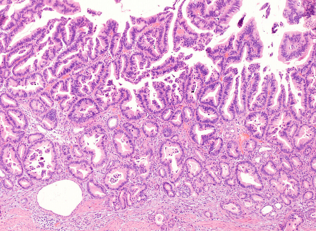
| Light micrograph of a haematoxylin and eosin-stained section from a gallbladder cancer. On the surface (top), the tumour has papillary and villous architecture with finger-like projections. The deeper aspects (bottom) show well-differentiated glands surrounded by fibrous stroma. | |
| Lizenzart: | Lizenzpflichtig |
| Credit: | Science Photo Library / WEBPATHOLOGY |
| Bildgröße: | 4096 px × 3000 px |
| Modell-Rechte: | nicht erforderlich |
| Eigentums-Rechte: | nicht erforderlich |
| Restrictions: | - |
Preise für dieses Bild ab 15 €
Universitäten & Organisationen
(Informationsmaterial Digital, Informationsmaterial Print, Lehrmaterial Digital etc.)
ab 15 €
Redaktionell
(Bücher, Bücher: Sach- und Fachliteratur, Digitale Medien (redaktionell) etc.)
ab 30 €
Werbung
(Anzeigen, Aussenwerbung, Digitale Medien, Fernsehwerbung, Karten, Werbemittel, Zeitschriften etc.)
ab 55 €
Handelsprodukte
(bedruckte Textilie, Kalender, Postkarte, Grußkarte, Verpackung etc.)
ab 75 €
Pauschalpreise
Rechtepakete für die unbeschränkte Bildnutzung in Print oder Online
ab 495 €
Keywords
- abnormal,
- Adenokarzinom,
- Diagnose,
- Galle,
- Gallenblase,
- gastrointestinal,
- Histologie,
- histologisch,
- Histopathologie,
- histopathologisch,
- Kondition,
- Krankheit,
- Krebs,
- krebsartig,
- lichtmikroskopische Aufnahme,
- maligne,
- Malignom,
- Medizin,
- medizinisch,
- Mikroskopie,
- Niemand,
- Onkologie,
- onkologisch,
- Organ,
- Pathologie,
- pathologisch,
- Störung,
- ungesund,
- Verdauungssystem