Tuberculosis of the lung, illustration
Bildnummer 14077910
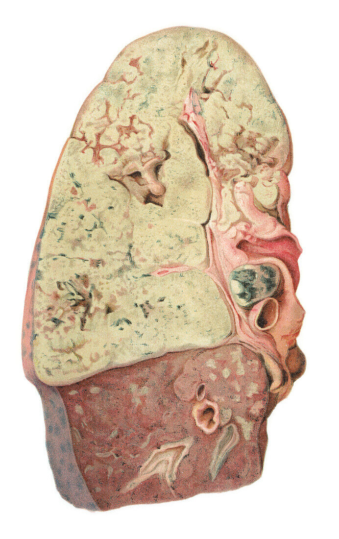
| Tuberculosis of the lung, illustration. Tuberculosis infection of the lung in which the upper lobe presents with a firm, dense consistency, and the tissue is largely devoid of airways. The yellowish-colour cut surface is friable and crumbly representing confluent necrotic cellular debris. Infected lymphoid tissue is noted at the root of the lung. Small foci of infection occur in the lower lobe. Tuberculosis bacteria are resistant to the tissue macrophages that attempt to destroy them and will replicate inside macrophages eventually killing these cells resulting in granulomas and necrosis. From Bollinger, O. 1901 Atlas und Gundriss der Pathologischen Anatomie, vol 1. Lehmann, Munich. | |
| Lizenzart: | Lizenzpflichtig |
| Credit: | Science Photo Library / Microscape |
| Bildgröße: | 3270 px × 5197 px |
| Modell-Rechte: | nicht erforderlich |
| Eigentums-Rechte: | nicht erforderlich |
| Restrictions: | - |
Preise für dieses Bild ab 15 €
Universitäten & Organisationen
(Informationsmaterial Digital, Informationsmaterial Print, Lehrmaterial Digital etc.)
ab 15 €
Redaktionell
(Bücher, Bücher: Sach- und Fachliteratur, Digitale Medien (redaktionell) etc.)
ab 30 €
Werbung
(Anzeigen, Aussenwerbung, Digitale Medien, Fernsehwerbung, Karten, Werbemittel, Zeitschriften etc.)
ab 55 €
Handelsprodukte
(bedruckte Textilie, Kalender, Postkarte, Grußkarte, Verpackung etc.)
ab 75 €
Pauschalpreise
Rechtepakete für die unbeschränkte Bildnutzung in Print oder Online
ab 495 €