Brain tumour, DTI MRI scans
Bildnummer 13950253
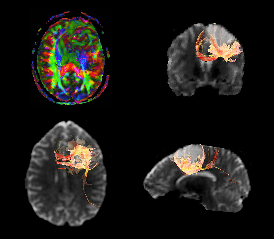
| Coloured diffusion tensor imaging (DTI) magnetic resonance imaging (MRI) scans of nerve pathways (red, blue and green) in the brain of a 20 year old male patient with a tumour (light grey) in the left temporoparietal region. Diffusion tensor imaging measures the direction of water diffusion, which in the brain reveals the orientation of nerve fibres. At top left, red fibres have a left to right orientation, green a front to back orientation and blue an up and down orientation. The tumour interrupts the course of the pyramidal tracts (red) in the left hemisphere. The pyramidal tracts are responsible for voluntary control of the muscles. | |
| Lizenzart: | Lizenzpflichtig |
| Credit: | Science Photo Library / Zephyr |
| Bildgröße: | 3969 px × 3455 px |
| Modell-Rechte: | nicht erforderlich |
| Eigentums-Rechte: | nicht erforderlich |
| Restrictions: | - |
Preise für dieses Bild ab 15 €
Universitäten & Organisationen
(Informationsmaterial Digital, Informationsmaterial Print, Lehrmaterial Digital etc.)
ab 15 €
Redaktionell
(Bücher, Bücher: Sach- und Fachliteratur, Digitale Medien (redaktionell) etc.)
ab 30 €
Werbung
(Anzeigen, Aussenwerbung, Digitale Medien, Fernsehwerbung, Karten, Werbemittel, Zeitschriften etc.)
ab 55 €
Handelsprodukte
(bedruckte Textilie, Kalender, Postkarte, Grußkarte, Verpackung etc.)
ab 75 €
Pauschalpreise
Rechtepakete für die unbeschränkte Bildnutzung in Print oder Online
ab 495 €
Keywords
- 20er Jahre,
- 3D,
- abnormal,
- axial,
- Ballaststoff,
- Diagnose,
- Dreidimensional,
- dti,
- dti Scan,
- Erwachsene,
- farbig,
- Fasern,
- Frontal,
- geduldig,
- Gehirn,
- Hirnscan,
- Jung,
- Kondition,
- Krankheit,
- Magnetresonanztomografie,
- Mann,
- Männlich,
- Medizin,
- medizinisch,
- menschlicher Körper,
- MRT-Untersuchung,
- Nerven,
- Nerventrakt,
- Neuroimaging,
- Niemand,
- Profil,
- Scan,
- schwarzer Hintergrund,
- Seitenansicht,
- Störung,
- ungesund,
- Weg,
- Wege,
- weiße Substanz,
- Wunde,
- zentrales Nervensystem,
- zwanzig,
- zwanziger Jahre