Brain lesion, DTI MRI scan
Bildnummer 13950250
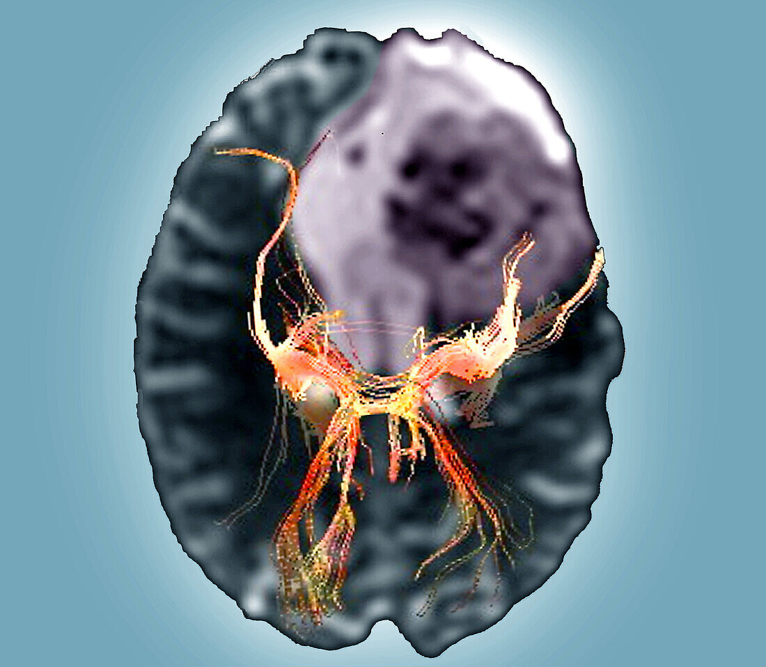
| Coloured diffusion tensor imaging (DTI) magnetic resonance imaging (MRI) scan of nerve pathways (orange) in the brain of a 44 year old male patient with a large lesion (purple) in the left temporoparietal region. The lesion is probably a glioblastoma, a malignant (cancerous) tumour that arises from the supporting glial cells in the brain. Diffusion tensor imaging measures the direction of water diffusion, which in the brain reveals the orientation of nerve fibres. This scan shows that some of the fibres have been displaced by the lesion. | |
| Lizenzart: | Lizenzpflichtig |
| Credit: | Science Photo Library / Zephyr |
| Bildgröße: | 3969 px × 3455 px |
| Modell-Rechte: | nicht erforderlich |
| Eigentums-Rechte: | nicht erforderlich |
| Restrictions: | - |
Preise für dieses Bild ab 15 €
Universitäten & Organisationen
(Informationsmaterial Digital, Informationsmaterial Print, Lehrmaterial Digital etc.)
ab 15 €
Redaktionell
(Bücher, Bücher: Sach- und Fachliteratur, Digitale Medien (redaktionell) etc.)
ab 30 €
Werbung
(Anzeigen, Aussenwerbung, Digitale Medien, Fernsehwerbung, Karten, Werbemittel, Zeitschriften etc.)
ab 55 €
Handelsprodukte
(bedruckte Textilie, Kalender, Postkarte, Grußkarte, Verpackung etc.)
ab 75 €
Pauschalpreise
Rechtepakete für die unbeschränkte Bildnutzung in Print oder Online
ab 495 €
Keywords
- 3D,
- 40er Jahre,
- abnormal,
- axial,
- Ballaststoff,
- Blauer Hintergrund,
- Diagnose,
- Dreidimensional,
- dti,
- dti Scan,
- Erwachsene,
- farbig,
- Fasern,
- geduldig,
- Gehirn,
- Hirnscan,
- Kondition,
- Krankheit,
- Magnetresonanztomografie,
- Mann,
- Männlich,
- Medizin,
- medizinisch,
- menschlicher Körper,
- mittleren Alters,
- MRT-Untersuchung,
- Nerven,
- Nerventrakt,
- Neuroimaging,
- Niemand,
- Störung,
- ungesund,
- Vierziger Jahre,
- Weg,
- Wege,
- weiße Substanz,
- Wunde,
- zentrales Nervensystem