Developing tooth, SEM
Bildnummer 13838907
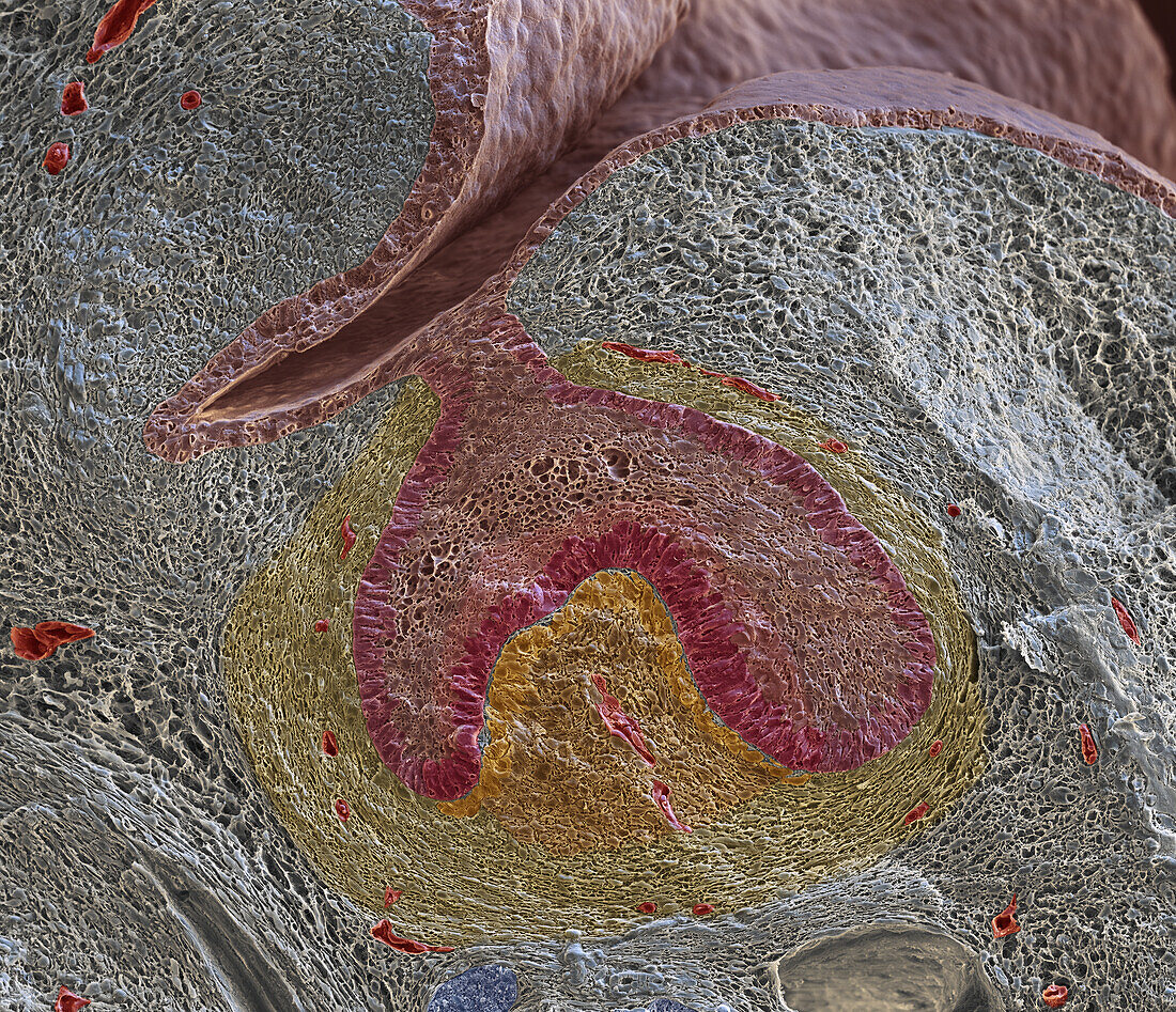
| Coloured scanning electron micrograph (SEM) of a freeze-fracture through the jaw of a 16.5 day old mouse embryo showing the late cap stage of a developing molar tooth. The gums are the thin layer (pink) at top, with fibroblasts (collagen-producing cells) and connective tissue (grey) below them. The dental follicle (yellow) contains pre-odontoblasts (orange) and pre-ameloblasts (red). Ameloblasts are the cells that secrete the enamel that covers the crown (visible part) of the tooth. Odontoblasts create dentine, the mineralised connective tissue that forms the bulk of the tooth. The picture was created as part of a cooperation with Quintessenz Publishing. Magnification: x300 when printed at 15cm wide. | |
| Lizenzart: | Lizenzpflichtig |
| Credit: | Science Photo Library / EYE OF SCIENCE |
| Bildgröße: | 4096 px × 3527 px |
| Modell-Rechte: | nicht erforderlich |
| Eigentums-Rechte: | nicht erforderlich |
| Restrictions: |
|
Preise für dieses Bild ab 15 €
Universitäten & Organisationen
(Informationsmaterial Digital, Informationsmaterial Print, Lehrmaterial Digital etc.)
ab 15 €
Redaktionell
(Bücher, Bücher: Sach- und Fachliteratur, Digitale Medien (redaktionell) etc.)
ab 30 €
Werbung
(Anzeigen, Aussenwerbung, Digitale Medien, Fernsehwerbung, Karten, Werbemittel, Zeitschriften etc.)
ab 55 €
Handelsprodukte
(bedruckte Textilie, Kalender, Postkarte, Grußkarte, Verpackung etc.)
ab 75 €
Pauschalpreise
Rechtepakete für die unbeschränkte Bildnutzung in Print oder Online
ab 495 €
Keywords
- Anatomie,
- anatomisch,
- Bildung,
- Biologie,
- biologisch,
- dental,
- Dentin,
- Emaille,
- Embryo,
- Embryonal,
- Entwicklung,
- farbig,
- Formation,
- gefärbt,
- Gefrierbruch,
- gefriergebrochen,
- gesund,
- Gewebe,
- Histologie,
- histologisch,
- Kappenstadium,
- Kauen,
- Kiefer,
- Mikroskopie,
- Mineral,
- Mund,
- Niemand,
- normal,
- Odontoblasten,
- Prä-Ameloblasten,
- Prä-Odontoblasten,
- Produktion,
- rasterelektronenmikroskopische Aufnahme,
- REM,
- Zahn,
- Zähne,
- Zahnentwicklung,
- Zahnheilkunde,
- Zelle,
- Zellen