Cells and bacteria from gingival sulcus, SEM
Bildnummer 13838901
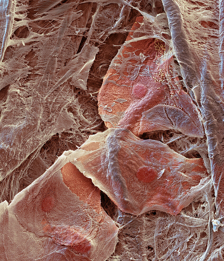
| Coloured scanning electron micrograph (SEM) of non-keratinised epithelial cells (pink) and bacteria (yellow) collected from a healthy gingival sulcus using a paper point. Fibres (grey-brown) from the paper are visible in the background. The gingival sulcus is the area where the gums and teeth meet. Only a few bacteria are present indicating that the sulcus was normally colonised. The picture was created as part of a cooperation with Quintessenz Publishing. Magnification: x1, 200 when printed at 15cm wide. | |
| Lizenzart: | Lizenzpflichtig |
| Credit: | Science Photo Library / EYE OF SCIENCE |
| Bildgröße: | 3516 px × 4096 px |
| Modell-Rechte: | nicht erforderlich |
| Eigentums-Rechte: | nicht erforderlich |
| Restrictions: |
|
Preise für dieses Bild ab 15 €
Universitäten & Organisationen
(Informationsmaterial Digital, Informationsmaterial Print, Lehrmaterial Digital etc.)
ab 15 €
Redaktionell
(Bücher, Bücher: Sach- und Fachliteratur, Digitale Medien (redaktionell) etc.)
ab 30 €
Werbung
(Anzeigen, Aussenwerbung, Digitale Medien, Fernsehwerbung, Karten, Werbemittel, Zeitschriften etc.)
ab 55 €
Handelsprodukte
(bedruckte Textilie, Kalender, Postkarte, Grußkarte, Verpackung etc.)
ab 75 €
Pauschalpreise
Rechtepakete für die unbeschränkte Bildnutzung in Print oder Online
ab 495 €
Keywords
- bakteriell,
- Bakterien,
- Bakterium,
- Biologie,
- biologisch,
- dental,
- eingefärbt,
- Epithel,
- epithelial,
- Epithelien,
- Epithelzellen,
- Farbig,
- Fasern,
- gefärbt,
- Gesund,
- Gingiva,
- Histologie,
- histologisch,
- Kauen,
- Kaugummi,
- Mikroskopie,
- Mund,
- Mundschleimhaut,
- Niemand,
- normal,
- Papier,
- rasterelektronenmikroskopische Aufnahme,
- REM,
- Zahn,
- Zähne,
- Zahnheilkunde,
- Zahntasche,
- Zelle,
- Zellen