Tooth dentine-pulp boundary, SEM
Bildnummer 13838849
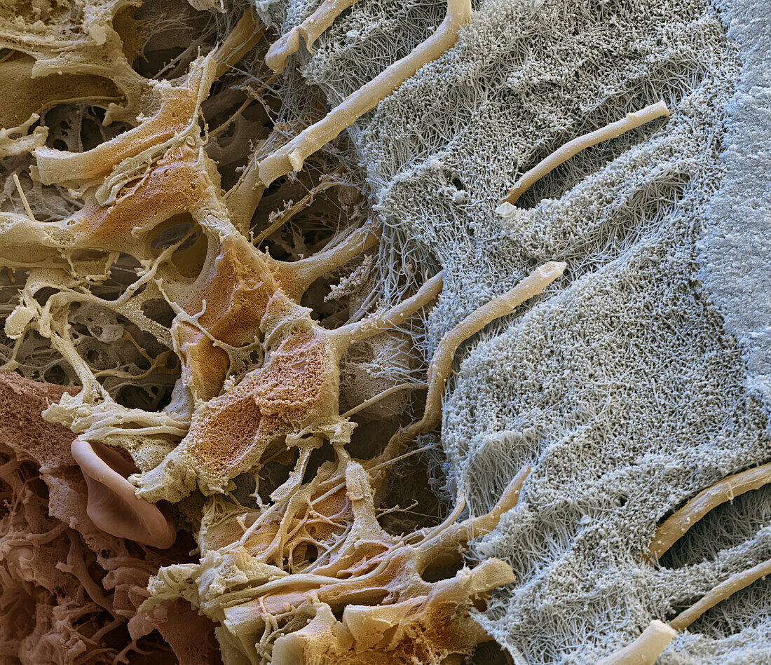
| Coloured scanning electron micrograph (SEM) of a freeze-fracture through a human tooth. At far right is the dentine (blue), the mineralised connective tissue that forms the bulk of the tooth. To its left is a dense network of collagen known as predentine (grey), which will calcify into dentine. Within the dentine are dentine tubules, which have been formed by the cytoplasmic extensions (beige) of odontoblast cells (dentine-producing cells). The odontoblast cells originate in the pulp (red, bottom left) and allow the dentine to rebuild itself. The picture was created as part of a cooperation with Quintessenz Publishing. Magnification: x4, 500 when printed at 15cm wide. | |
| Lizenzart: | Lizenzpflichtig |
| Credit: | Science Photo Library / EYE OF SCIENCE |
| Bildgröße: | 4096 px × 3534 px |
| Modell-Rechte: | nicht erforderlich |
| Eigentums-Rechte: | nicht erforderlich |
| Restrictions: |
|
Preise für dieses Bild ab 15 €
Universitäten & Organisationen
(Informationsmaterial Digital, Informationsmaterial Print, Lehrmaterial Digital etc.)
ab 15 €
Redaktionell
(Bücher, Bücher: Sach- und Fachliteratur, Digitale Medien (redaktionell) etc.)
ab 30 €
Werbung
(Anzeigen, Aussenwerbung, Digitale Medien, Fernsehwerbung, Karten, Werbemittel, Zeitschriften etc.)
ab 55 €
Handelsprodukte
(bedruckte Textilie, Kalender, Postkarte, Grußkarte, Verpackung etc.)
ab 75 €
Pauschalpreise
Rechtepakete für die unbeschränkte Bildnutzung in Print oder Online
ab 495 €
Keywords
- Anatomie,
- anatomisch,
- Biologie,
- biologisch,
- dental,
- Dentin,
- Dentintubuli,
- Farbig,
- gefriergebrochen,
- Histologie,
- histologisch,
- Kanal,
- Kanäle,
- Kauen,
- Kollagen,
- Mikroskopie,
- Mund,
- Niemand,
- Odontoblasten,
- Prädentin,
- rasterelektronenmikroskopische Aufnahme,
- REM,
- Sektion,
- sektioniert,
- Struktur,
- Zahn,
- Zähne,
- Zahnheilkunde