Developing tooth, SEM
Bildnummer 13838547
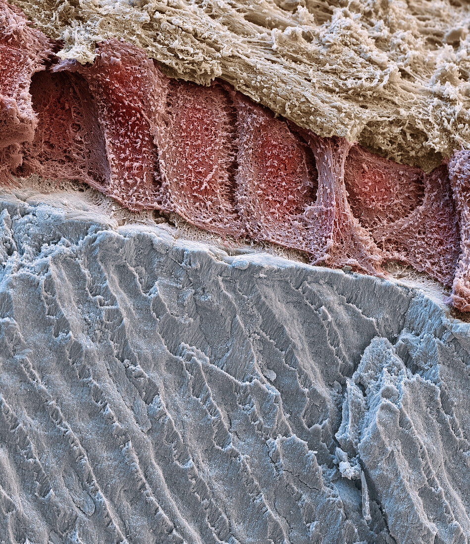
| Coloured scanning electron micrograph (SEM) of a developing tooth. At bottom is the enamel (grey) of the tooth, which is secreted by ameloblast cells (red). These cells are initially long and cylindrical but shorten during the growth of the tooth. They die off before the tooth erupts. Across top are cells (beige) of the stratum intermedium, an epithelial tissue associated with the ameloblasts. The picture was created as part of a cooperation with Quintessenz Publishing. Magnification: x2000 when printed at 15cm wide. | |
| Lizenzart: | Lizenzpflichtig |
| Credit: | Science Photo Library / EYE OF SCIENCE |
| Bildgröße: | 3533 px × 4096 px |
| Modell-Rechte: | nicht erforderlich |
| Eigentums-Rechte: | nicht erforderlich |
| Restrictions: |
|
Preise für dieses Bild ab 15 €
Universitäten & Organisationen
(Informationsmaterial Digital, Informationsmaterial Print, Lehrmaterial Digital etc.)
ab 15 €
Redaktionell
(Bücher, Bücher: Sach- und Fachliteratur, Digitale Medien (redaktionell) etc.)
ab 30 €
Werbung
(Anzeigen, Aussenwerbung, Digitale Medien, Fernsehwerbung, Karten, Werbemittel, Zeitschriften etc.)
ab 55 €
Handelsprodukte
(bedruckte Textilie, Kalender, Postkarte, Grußkarte, Verpackung etc.)
ab 75 €
Pauschalpreise
Rechtepakete für die unbeschränkte Bildnutzung in Print oder Online
ab 495 €
Keywords
- Ameloblast,
- Ameloblasten,
- Anatomie,
- anatomisch,
- Bildung,
- Biologie,
- biologisch,
- dental,
- Emaille,
- Embryonaler Zahn,
- Entwicklung,
- Epithel,
- epithelial,
- farbig,
- Formation,
- gefärbt,
- gesund,
- Gewebe,
- Histologie,
- histologisch,
- Kauen,
- Kiefer,
- Mikroskopie,
- Mineral,
- Mund,
- Niemand,
- normal,
- Produktion,
- rasterelektronenmikroskopische Aufnahme,
- REM,
- Schmelz,
- Stratium Intermedium,
- Zahn,
- Zähne,
- Zahnentwicklung,
- Zahnheilkunde,
- Zahnschmelz,
- Zelle,
- Zellen