Chondrosarcoma, light micrograph
Bildnummer 13765412
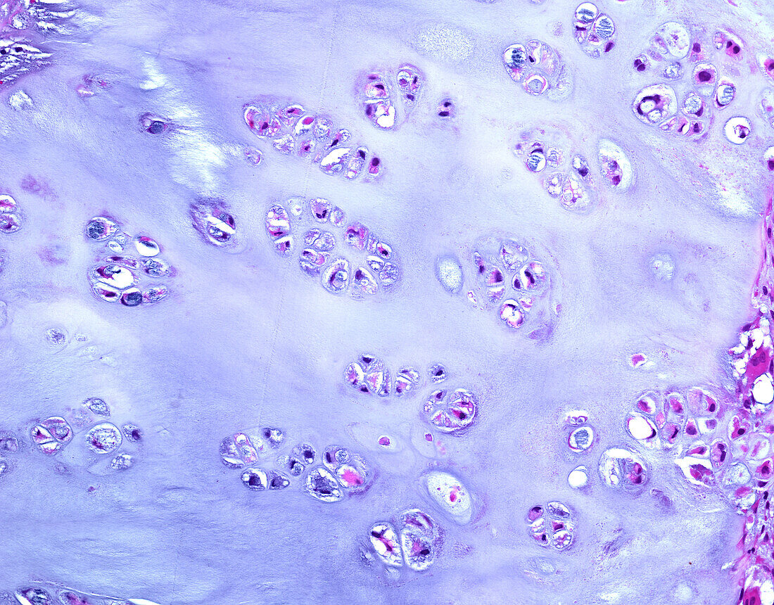
| Light micrograph of a section through a low-grade (Grade 1) chondrosarcoma. The nuclei of chondrocytes (tumour cells) are arranged in clusters in a background of bluish cartilaginous matrix. The nuclei lack significant cytologic atypia. Chondrosarcoma is a malignant tumour which shows chondroid (cartilaginous) differentiation. It is a tumour of adulthood and old age, most commonly presenting in 4th to 6th decades of life. The most common locations are the pelvic bones, ribs, shoulder girdle and the femur (thigh bone). Haematoxylin and eosin stain. | |
| Lizenzart: | Lizenzpflichtig |
| Credit: | Science Photo Library / WEBPATHOLOGY |
| Bildgröße: | 4094 px × 3200 px |
| Modell-Rechte: | nicht erforderlich |
| Eigentums-Rechte: | nicht erforderlich |
| Restrictions: | - |
Preise für dieses Bild ab 15 €
Universitäten & Organisationen
(Informationsmaterial Digital, Informationsmaterial Print, Lehrmaterial Digital etc.)
ab 15 €
Redaktionell
(Bücher, Bücher: Sach- und Fachliteratur, Digitale Medien (redaktionell) etc.)
ab 30 €
Werbung
(Anzeigen, Aussenwerbung, Digitale Medien, Fernsehwerbung, Karten, Werbemittel, Zeitschriften etc.)
ab 55 €
Handelsprodukte
(bedruckte Textilie, Kalender, Postkarte, Grußkarte, Verpackung etc.)
ab 75 €
Pauschalpreise
Rechtepakete für die unbeschränkte Bildnutzung in Print oder Online
ab 495 €