Madurella mycetomatis fungus, light micrograph
Bildnummer 13765193
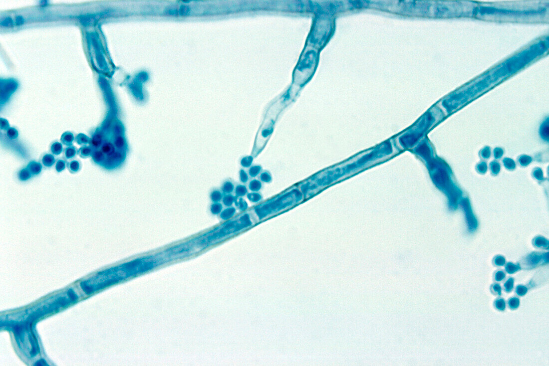
| Light micrograph of the fungus Madurella mycetomatis showing conidiophores (branches with terminal clusters of ovals) and septate hyphae (one diagonally across centre). Hyphae are the branching, filamentous structures of a fungus, that in this species are separated by septa (walls) into cellular compartments. Conidiophores are specialised hyphae that produce asexual spores (reproductive cells, ovals) called conidia. M. mycetomatis can cause an infection of the subcutaneous tissue (deepest layer of skin) known as mycetoma. | |
| Lizenzart: | Lizenzpflichtig |
| Credit: | Science Photo Library / CDC |
| Bildgröße: | 3620 px × 2414 px |
| Modell-Rechte: | nicht erforderlich |
| Eigentums-Rechte: | nicht erforderlich |
| Restrictions: | - |
Preise für dieses Bild ab 15 €
Universitäten & Organisationen
(Informationsmaterial Digital, Informationsmaterial Print, Lehrmaterial Digital etc.)
ab 15 €
Redaktionell
(Bücher, Bücher: Sach- und Fachliteratur, Digitale Medien (redaktionell) etc.)
ab 30 €
Werbung
(Anzeigen, Aussenwerbung, Digitale Medien, Fernsehwerbung, Karten, Werbemittel, Zeitschriften etc.)
ab 55 €
Handelsprodukte
(bedruckte Textilie, Kalender, Postkarte, Grußkarte, Verpackung etc.)
ab 75 €
Pauschalpreise
Rechtepakete für die unbeschränkte Bildnutzung in Print oder Online
ab 495 €