Pine needle.
Bildnummer 13756290
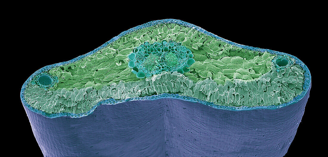
| Pine needle. Coloured scanning electron micrograph (SEM) of a freeze-fracture through a leaf (needle) of a pine tree (Pinus sp.). The leaves are needle-like in order to present a large surface area for photosynthesis but prevent too much water loss (transpiration). They have an epidermis of thick walled cells covered with a thick layer of cuticle. The mesophyll layer under the epidermis is made up of parenchyma cells. The vascular cylinder (centre) is surrounded by the endodermis which regulates water and mineral movement. The centre of the needle is occupied by two vascular bundles, each one made up of xylem and phloem tissue. These are surrounded by a thick pericycle (large-celled region) and a layer of endodermis (necklace-like ring of large cells). A resin duct is seen at either side of the section. Magnification: x46 when printed 10 centimetres wide. | |
| Lizenzart: | Lizenzpflichtig |
| Credit: | Science Photo Library / Gschmeissner, Steve |
| Bildgröße: | 6096 px × 2923 px |
| Modell-Rechte: | nicht erforderlich |
| Restrictions: | - |
Preise für dieses Bild ab 15 €
Universitäten & Organisationen
(Informationsmaterial Digital, Informationsmaterial Print, Lehrmaterial Digital etc.)
ab 15 €
Redaktionell
(Bücher, Bücher: Sach- und Fachliteratur, Digitale Medien (redaktionell) etc.)
ab 30 €
Werbung
(Anzeigen, Aussenwerbung, Digitale Medien, Fernsehwerbung, Karten, Werbemittel, Zeitschriften etc.)
ab 55 €
Handelsprodukte
(bedruckte Textilie, Kalender, Postkarte, Grußkarte, Verpackung etc.)
ab 75 €
Pauschalpreise
Rechtepakete für die unbeschränkte Bildnutzung in Print oder Online
ab 495 €