Striated muscle fibre, light micrograph
Bildnummer 13756184
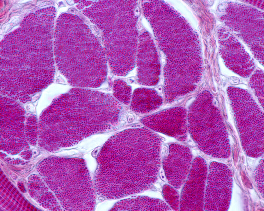
| Light micrograph of a cross-sectioned skeletal muscle stained with Mallory's phosphotungstic acid haematoxylin (PTAH). Each oval or polygonal structure corresponds to a cross-sectioned myocyte. Inside each one there is a dotted pattern that occupies the entire cytoplasm that corresponds to the set of myofibrils. The nuclei, which are poorly stained with this technique, are displaced to the periphery. The narrow space between fibres is the endomysium, where blood capillaries are found. | |
| Lizenzart: | Lizenzpflichtig |
| Credit: | Science Photo Library / JOSE CALVO |
| Bildgröße: | 3840 px × 3072 px |
| Modell-Rechte: | nicht erforderlich |
| Eigentums-Rechte: | nicht erforderlich |
| Restrictions: | - |
Preise für dieses Bild ab 15 €
Universitäten & Organisationen
(Informationsmaterial Digital, Informationsmaterial Print, Lehrmaterial Digital etc.)
ab 15 €
Redaktionell
(Bücher, Bücher: Sach- und Fachliteratur, Digitale Medien (redaktionell) etc.)
ab 30 €
Werbung
(Anzeigen, Aussenwerbung, Digitale Medien, Fernsehwerbung, Karten, Werbemittel, Zeitschriften etc.)
ab 55 €
Handelsprodukte
(bedruckte Textilie, Kalender, Postkarte, Grußkarte, Verpackung etc.)
ab 75 €
Pauschalpreise
Rechtepakete für die unbeschränkte Bildnutzung in Print oder Online
ab 495 €