Mitosis, light micrograph
Bildnummer 13755892
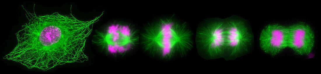
| Fluorescent light micrograph of cells during mitosis (nuclear division). Mitosis is the formation of two daughter nuclei from one parent nucleus. Fluorescent markers have been used to highlight DNA (deoxyribonucleic acid, pink) and alpha tubulin (green), a component of microtubules. The cell at left is in prophase, the nuclear envelope is dissolving and the chromosomes are condensing. The cell progress through prometaphase (second from left) to metaphase (centre), where the chromosomes align along the centre of the cell. The chromosomes start to move to the opposite poles, guided by microtubules, during anaphase (second from right). The cell at right is in telophase, when the separated chromosomes have moved to opposite ends of the cell and two new nuclei form around them. | |
| Lizenzart: | Lizenzpflichtig |
| Credit: | Science Photo Library / DR. JUAN F. GIMENEZ-ABIAN |
| Bildgröße: | 6209 px × 1429 px |
| Modell-Rechte: | nicht erforderlich |
| Eigentums-Rechte: | nicht erforderlich |
| Restrictions: | - |
Preise für dieses Bild ab 15 €
Universitäten & Organisationen
(Informationsmaterial Digital, Informationsmaterial Print, Lehrmaterial Digital etc.)
ab 15 €
Redaktionell
(Bücher, Bücher: Sach- und Fachliteratur, Digitale Medien (redaktionell) etc.)
ab 30 €
Werbung
(Anzeigen, Aussenwerbung, Digitale Medien, Fernsehwerbung, Karten, Werbemittel, Zeitschriften etc.)
ab 55 €
Handelsprodukte
(bedruckte Textilie, Kalender, Postkarte, Grußkarte, Verpackung etc.)
ab 75 €
Pauschalpreise
Rechtepakete für die unbeschränkte Bildnutzung in Print oder Online
ab 495 €
Keywords
- alpha-Tubulin,
- Biologie,
- biologisch,
- Chromosom,
- Chromosomen,
- Desoxiribonukleinsäure,
- DNA,
- Einteilung,
- Fluoreszenz,
- fluoreszierend,
- Genetik,
- kopierend,
- lichtmikroskopische Aufnahme,
- Mikroskopie,
- Mitose,
- mitotische Spindel,
- Niemand,
- Prometaphase,
- Reproduktion,
- schwarzer Hintergrund,
- Separieren,
- Teilen,
- Wachstum,
- Zellbilogie,
- Zelle,
- Zellen,
- Zytologie,
- Zytologisch,
- Zytoskelett