Mitosis, light micrograph
Bildnummer 13755838
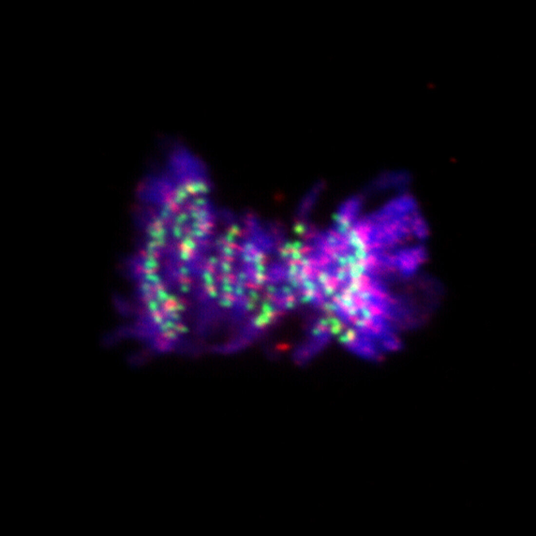
| Fluorescent light micrograph of a cell during the metaphase stage of mitosis (nuclear division). Mitosis is the formation of two daughter nuclei from one parent nucleus. Fluorescent markers have been used to highlight DNA (deoxyribonucleic acid, blue), cohesin (red) and kinetochores (green). Cohesin hold sister chromatids together. During metaphase the chromosomes align along the centre of the cell. In the next stage the sister chromatids of each chromosome are pulled to opposite poles by the mitotic spindle, which attach to the chromosomes via the kinetochores. | |
| Lizenzart: | Lizenzpflichtig |
| Credit: | Science Photo Library / DR. JUAN F. GIMENEZ-ABIAN |
| Bildgröße: | 2963 px × 2963 px |
| Modell-Rechte: | nicht erforderlich |
| Eigentums-Rechte: | nicht erforderlich |
| Restrictions: | - |
Preise für dieses Bild ab 15 €
Universitäten & Organisationen
(Informationsmaterial Digital, Informationsmaterial Print, Lehrmaterial Digital etc.)
ab 15 €
Redaktionell
(Bücher, Bücher: Sach- und Fachliteratur, Digitale Medien (redaktionell) etc.)
ab 30 €
Werbung
(Anzeigen, Aussenwerbung, Digitale Medien, Fernsehwerbung, Karten, Werbemittel, Zeitschriften etc.)
ab 55 €
Handelsprodukte
(bedruckte Textilie, Kalender, Postkarte, Grußkarte, Verpackung etc.)
ab 75 €
Pauschalpreise
Rechtepakete für die unbeschränkte Bildnutzung in Print oder Online
ab 495 €
Keywords
- Biologie,
- biologisch,
- Chromosom,
- Chromosomen,
- Desoxiribonukleinsäure,
- DNA,
- Einteilung,
- Fluoreszenz,
- fluoreszierend,
- Genetik,
- kopierend,
- lichtmikroskopische Aufnahme,
- Mikroskopie,
- Mitose,
- Niemand,
- Reproduktion,
- schwarzer Hintergrund,
- Teilen,
- Wachstum,
- Zellbilogie,
- Zelle,
- Zellen,
- Zytologie,
- Zytologisch