Human chromosomes, light micrograph
Bildnummer 13755837
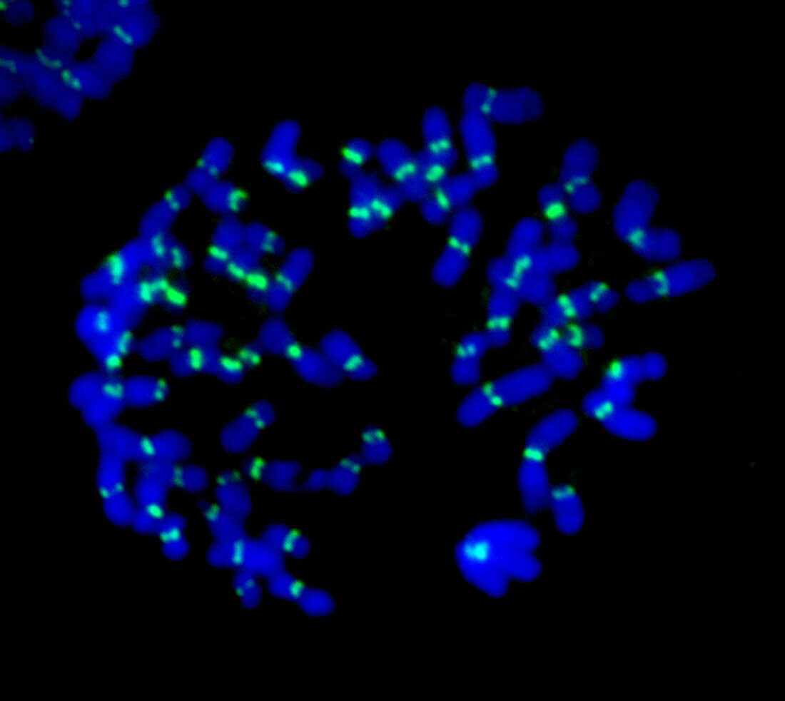
| Fluorescent light micrograph of human chromosomes. Fluorescent markers have been used to highlight DNA (deoxyribonucleic acid, blue) and kinetochores (green). During mitosis (nuclear division), the formation of two daughter nuclei from one parent nucleus, the spindle apparatus that separates' sister chromatids attaches to the chromosomes via the kinetochores. Two identical chromatids make up one chromosome, so each cell retains a copy of the parent cell's genetic information. | |
| Lizenzart: | Lizenzpflichtig |
| Credit: | Science Photo Library / DR. JUAN F. GIMENEZ-ABIAN |
| Bildgröße: | 3140 px × 2806 px |
| Modell-Rechte: | nicht erforderlich |
| Eigentums-Rechte: | nicht erforderlich |
| Restrictions: | - |
Preise für dieses Bild ab 15 €
Universitäten & Organisationen
(Informationsmaterial Digital, Informationsmaterial Print, Lehrmaterial Digital etc.)
ab 15 €
Redaktionell
(Bücher, Bücher: Sach- und Fachliteratur, Digitale Medien (redaktionell) etc.)
ab 30 €
Werbung
(Anzeigen, Aussenwerbung, Digitale Medien, Fernsehwerbung, Karten, Werbemittel, Zeitschriften etc.)
ab 55 €
Handelsprodukte
(bedruckte Textilie, Kalender, Postkarte, Grußkarte, Verpackung etc.)
ab 75 €
Pauschalpreise
Rechtepakete für die unbeschränkte Bildnutzung in Print oder Online
ab 495 €