Mitosis, light micrograph
Bildnummer 13755811
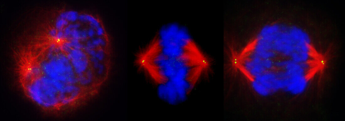
| Fluorescent light micrograph of cells during mitosis (nuclear division). Mitosis is the formation of two daughter nuclei from one parent nucleus. Fluorescent markers have been used to highlight DNA (deoxyribonucleic acid, blue) and alpha tubulin (red), a component of microtubules. Centrosomes appear as bright yellow dots. At left the cell is in prophase, the nuclear envelope dissolves and the chromosomes condense. The cell at centre is in metaphase, with the chromosomes aligned along the centre of the cell. The sister chromatids of each chromosome are pulled to opposite poles during anaphase (right). The chromatids are pulled apart by the mitotic spindle, the main component of which are microtubules. Each mitotic spindle emerges from a centrosome. | |
| Lizenzart: | Lizenzpflichtig |
| Credit: | Science Photo Library / DR. JUAN F. GIMENEZ-ABIAN |
| Bildgröße: | 5058 px × 1773 px |
| Modell-Rechte: | nicht erforderlich |
| Eigentums-Rechte: | nicht erforderlich |
| Restrictions: | - |
Preise für dieses Bild ab 15 €
Universitäten & Organisationen
(Informationsmaterial Digital, Informationsmaterial Print, Lehrmaterial Digital etc.)
ab 15 €
Redaktionell
(Bücher, Bücher: Sach- und Fachliteratur, Digitale Medien (redaktionell) etc.)
ab 30 €
Werbung
(Anzeigen, Aussenwerbung, Digitale Medien, Fernsehwerbung, Karten, Werbemittel, Zeitschriften etc.)
ab 55 €
Handelsprodukte
(bedruckte Textilie, Kalender, Postkarte, Grußkarte, Verpackung etc.)
ab 75 €
Pauschalpreise
Rechtepakete für die unbeschränkte Bildnutzung in Print oder Online
ab 495 €
Keywords
- alpha-Tubulin,
- Atomkern,
- Biologie,
- biologisch,
- Chromosom,
- Chromosomen,
- Desoxiribonukleinsäure,
- DNA,
- Einteilung,
- Fluoreszenz,
- fluoreszierend,
- Genetik,
- kopierend,
- lichtmikroskopische Aufnahme,
- Mikroskopie,
- Mitose,
- mitotische Spindel,
- Niemand,
- Reihenfolge,
- Reproduktion,
- schwarzer Hintergrund,
- Separieren,
- Teilen,
- Wachstum,
- Zellbilogie,
- Zelle,
- Zellen,
- Zytologie,
- Zytologisch,
- Zytoskelett