Simple columnar epithelium, light micrograph
Bildnummer 13742445
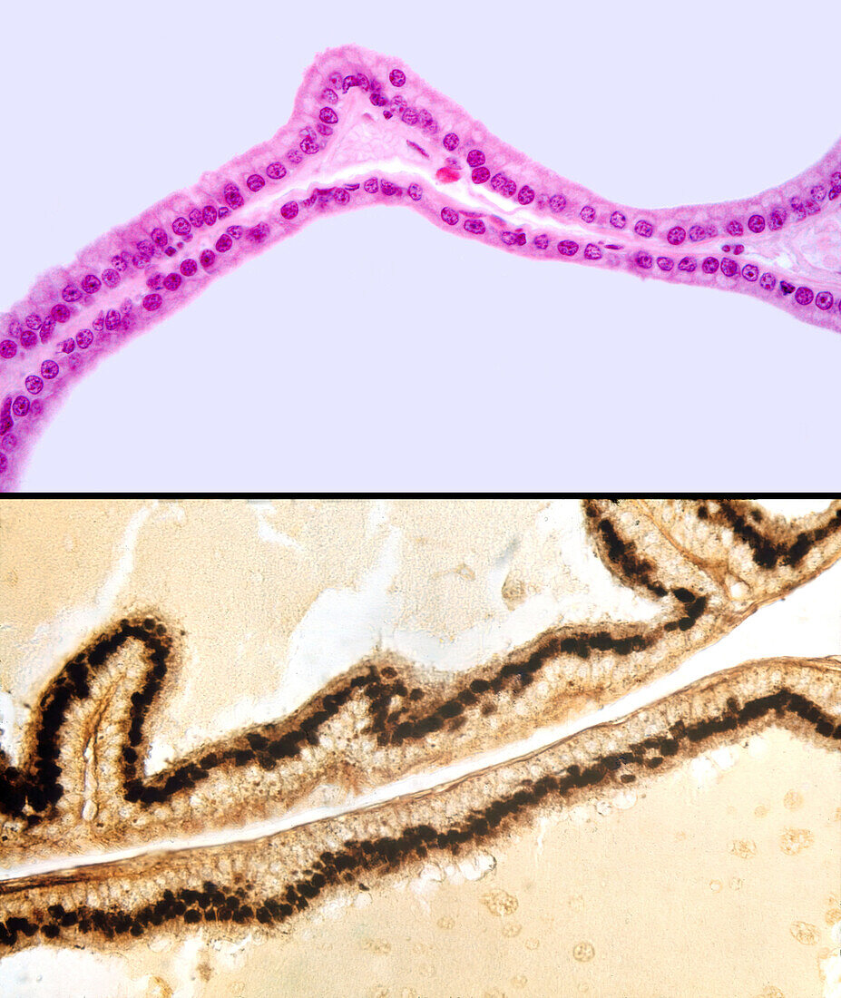
| Light micrograph of the septum between two alveoli of a rat coagulating gland lined by a simple columnar epithelium stained with haematoxylin and eosin (top) and the Da Fano silver method for Golgi apparatus (bottom). At top, epithelial cells show a supranuclear light band corresponding to the Golgi apparatus location (negative Golgi image). At bottom, these cells show a dark silver stained band above the nucleus corresponding to the location of the Golgi apparatus. | |
| Lizenzart: | Lizenzpflichtig |
| Credit: | Science Photo Library / JOSE CALVO |
| Bildgröße: | 3840 px × 4556 px |
| Modell-Rechte: | nicht erforderlich |
| Eigentums-Rechte: | nicht erforderlich |
| Restrictions: | - |
Preise für dieses Bild ab 15 €
Universitäten & Organisationen
(Informationsmaterial Digital, Informationsmaterial Print, Lehrmaterial Digital etc.)
ab 15 €
Redaktionell
(Bücher, Bücher: Sach- und Fachliteratur, Digitale Medien (redaktionell) etc.)
ab 30 €
Werbung
(Anzeigen, Aussenwerbung, Digitale Medien, Fernsehwerbung, Karten, Werbemittel, Zeitschriften etc.)
ab 55 €
Handelsprodukte
(bedruckte Textilie, Kalender, Postkarte, Grußkarte, Verpackung etc.)
ab 75 €
Pauschalpreise
Rechtepakete für die unbeschränkte Bildnutzung in Print oder Online
ab 495 €