Oxalate calcification in breast biopsy, light micrograph
Bildnummer 13733296
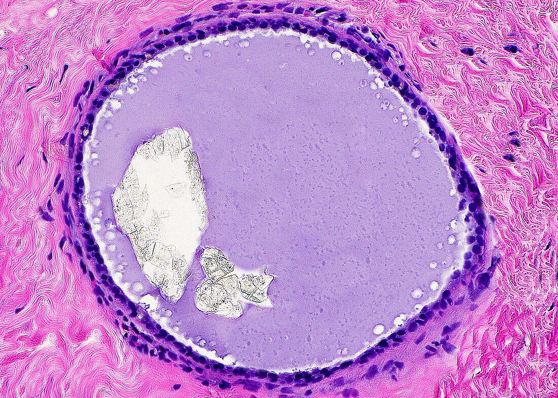
| Light micrograph of a breast microcyst (the circular blue structure) containing calcium oxalate crystals (the clear-yellowish irregular shaped structures within the light blue area). Calcifications such as these are often detected on mammography by a radiologist. The presence of the crystals is then confirmed by pathologists examining breast biopsy tissue to rule out breast cancer. Haematoxylin and eosin stained tissue section. Magnification: 200x when printed at 10 cm. | |
| Lizenzart: | Lizenzpflichtig |
| Credit: | Science Photo Library / ZIAD M. EL-ZAATARI |
| Bildgröße: | 5000 px × 3567 px |
| Modell-Rechte: | nicht erforderlich |
| Eigentums-Rechte: | nicht erforderlich |
| Restrictions: | - |
Preise für dieses Bild ab 15 €
Universitäten & Organisationen
(Informationsmaterial Digital, Informationsmaterial Print, Lehrmaterial Digital etc.)
ab 15 €
Redaktionell
(Bücher, Bücher: Sach- und Fachliteratur, Digitale Medien (redaktionell) etc.)
ab 30 €
Werbung
(Anzeigen, Aussenwerbung, Digitale Medien, Fernsehwerbung, Karten, Werbemittel, Zeitschriften etc.)
ab 55 €
Handelsprodukte
(bedruckte Textilie, Kalender, Postkarte, Grußkarte, Verpackung etc.)
ab 75 €
Pauschalpreise
Rechtepakete für die unbeschränkte Bildnutzung in Print oder Online
ab 495 €