Her2 protein, light micrograph
Bildnummer 13732458
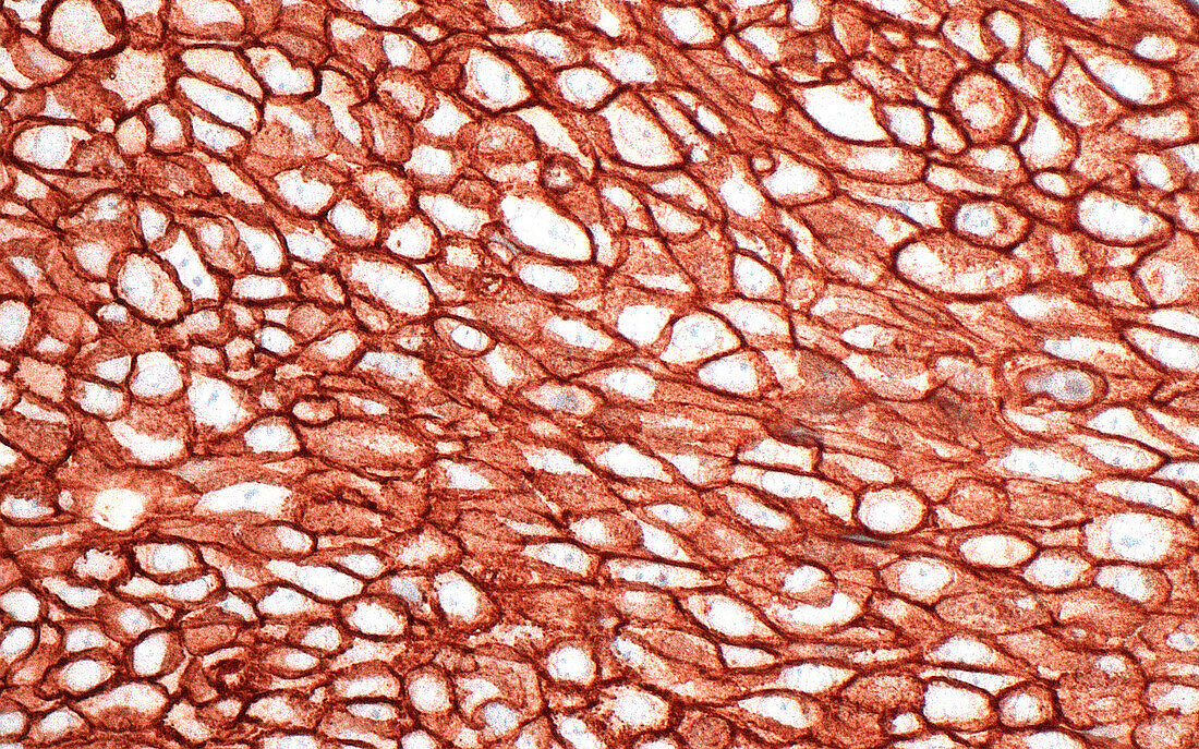
| Light micrograph of Her2 positive cells stained by immunohistochemistry. A pathologist can tell the stain is positive by its brown colour that highlights the contours of the individual cells. The presence of positive staining for Her2 can help guide therapy for cancer patients, such as those suffering from breast cancer. Haematoxylin and eosin stained tissue section. Magnification: 400x when printed at 10cm. | |
| Lizenzart: | Lizenzpflichtig |
| Credit: | Science Photo Library / ZIAD M. EL-ZAATARI |
| Bildgröße: | 5000 px × 3124 px |
| Modell-Rechte: | nicht erforderlich |
| Eigentums-Rechte: | nicht erforderlich |
| Restrictions: | - |
Preise für dieses Bild ab 15 €
Universitäten & Organisationen
(Informationsmaterial Digital, Informationsmaterial Print, Lehrmaterial Digital etc.)
ab 15 €
Redaktionell
(Bücher, Bücher: Sach- und Fachliteratur, Digitale Medien (redaktionell) etc.)
ab 30 €
Werbung
(Anzeigen, Aussenwerbung, Digitale Medien, Fernsehwerbung, Karten, Werbemittel, Zeitschriften etc.)
ab 55 €
Handelsprodukte
(bedruckte Textilie, Kalender, Postkarte, Grußkarte, Verpackung etc.)
ab 75 €
Pauschalpreise
Rechtepakete für die unbeschränkte Bildnutzung in Print oder Online
ab 495 €