Prostate intraepithelial neoplasia, light micrograph
Bildnummer 13732439
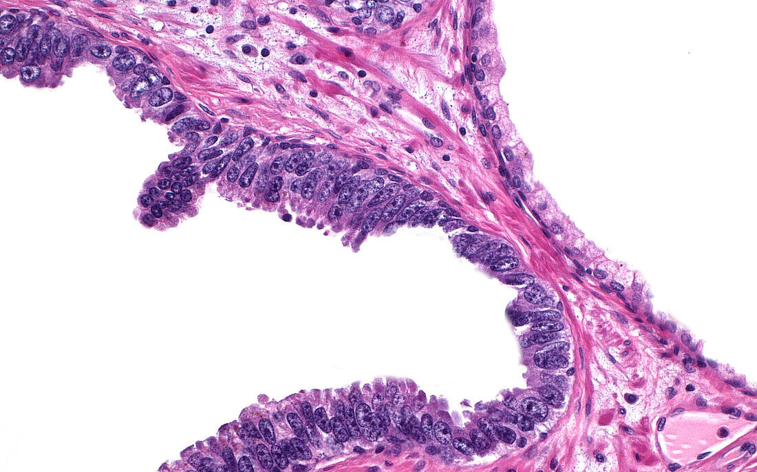
| Light micrograph of high grade prostatic intraepithelial neoplasia (PIN). On the left side is a prostate gland with hyperchromatic (dark blue) and enlarged lining cells. This change is termed intraepithelial neoplasia, and is seen more frequently in patients with prostate cancer. The gland on the right side of the picture is normal as seen by its lighter colour and smaller lining cells. Haematoxylin and eosin stained tissue section. Magnification: 200x when printed at 10 cm. | |
| Lizenzart: | Lizenzpflichtig |
| Credit: | Science Photo Library / ZIAD M. EL-ZAATARI |
| Bildgröße: | 5000 px × 3121 px |
| Modell-Rechte: | nicht erforderlich |
| Eigentums-Rechte: | nicht erforderlich |
| Restrictions: | - |
Preise für dieses Bild ab 15 €
Universitäten & Organisationen
(Informationsmaterial Digital, Informationsmaterial Print, Lehrmaterial Digital etc.)
ab 15 €
Redaktionell
(Bücher, Bücher: Sach- und Fachliteratur, Digitale Medien (redaktionell) etc.)
ab 30 €
Werbung
(Anzeigen, Aussenwerbung, Digitale Medien, Fernsehwerbung, Karten, Werbemittel, Zeitschriften etc.)
ab 55 €
Handelsprodukte
(bedruckte Textilie, Kalender, Postkarte, Grußkarte, Verpackung etc.)
ab 75 €
Pauschalpreise
Rechtepakete für die unbeschränkte Bildnutzung in Print oder Online
ab 495 €
Keywords
- Anatomie,
- Diagnose,
- Gewebe,
- Histologie,
- histologisch,
- Histopathologie,
- Kondition,
- Krankheit,
- lichtmikroskopische Aufnahme,
- männliches Fortpflanzungssystem,
- Medizin,
- menschlicher Körper,
- Mikroskopie,
- Pathologie,
- Prostata,
- Prostatakrebs,
- rutschen,
- Störung,
- Urologie,
- vergrößertes Bild,
- weißer Hintergrund,
- Zellen