Granulomas, light micrograph
Bildnummer 13732395
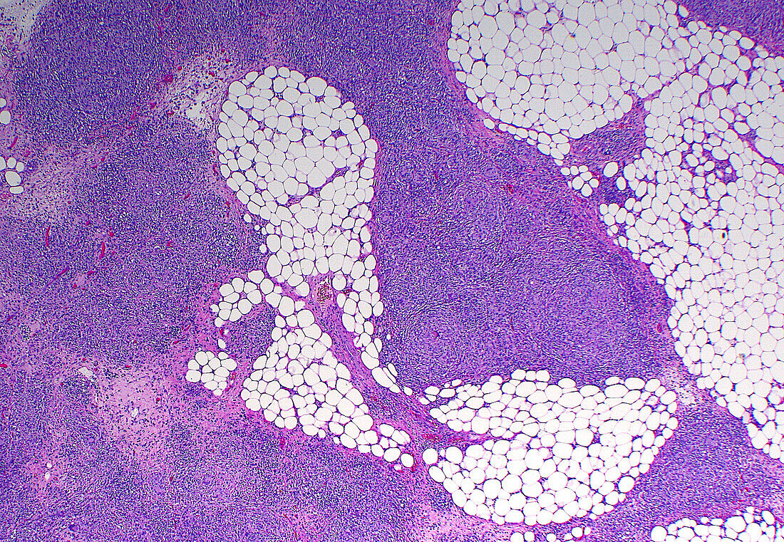
| Light micrograph of granulomas in fat. The granulomas are the blue areas of the image and are composed of histiocytes (a type of inflammatory cells). The white areas in between are fat cells. In this case, the granulomas formed in response to a fungal infection. Haematoxylin and eosin stained tissue section. Magnification: 20x when printed at 10 cm. | |
| Lizenzart: | Lizenzpflichtig |
| Credit: | Science Photo Library / ZIAD M. EL-ZAATARI |
| Bildgröße: | 5000 px × 3460 px |
| Modell-Rechte: | nicht erforderlich |
| Eigentums-Rechte: | nicht erforderlich |
| Restrictions: | - |
Preise für dieses Bild ab 15 €
Universitäten & Organisationen
(Informationsmaterial Digital, Informationsmaterial Print, Lehrmaterial Digital etc.)
ab 15 €
Redaktionell
(Bücher, Bücher: Sach- und Fachliteratur, Digitale Medien (redaktionell) etc.)
ab 30 €
Werbung
(Anzeigen, Aussenwerbung, Digitale Medien, Fernsehwerbung, Karten, Werbemittel, Zeitschriften etc.)
ab 55 €
Handelsprodukte
(bedruckte Textilie, Kalender, Postkarte, Grußkarte, Verpackung etc.)
ab 75 €
Pauschalpreise
Rechtepakete für die unbeschränkte Bildnutzung in Print oder Online
ab 495 €