Fat necrosis, light micrograph
Bildnummer 13732389
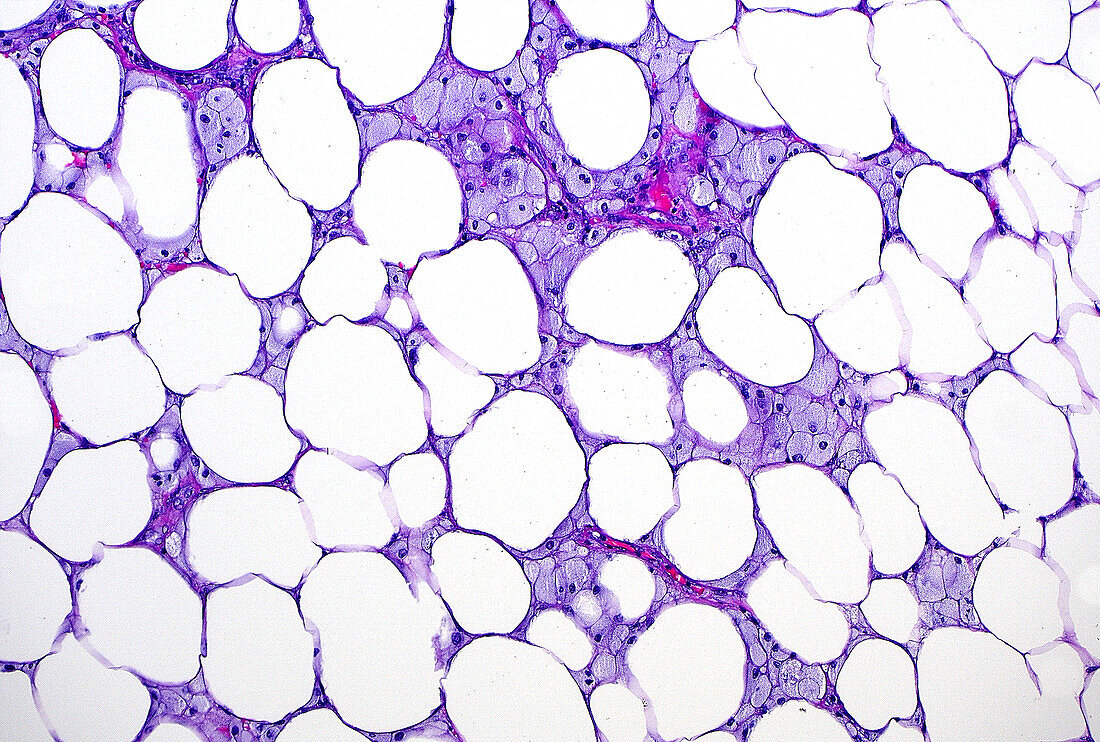
| Light micrograph of fat necrosis. Dying fat cells (the white circular spaces) have no nuclei and are admixed with foamy histiocytes (the blue-gray cells in between). Haematoxylin and eosin stained tissue section. Magnification: 100x when printed at 10 cm. | |
| Lizenzart: | Lizenzpflichtig |
| Credit: | Science Photo Library / ZIAD M. EL-ZAATARI |
| Bildgröße: | 5000 px × 3373 px |
| Modell-Rechte: | nicht erforderlich |
| Eigentums-Rechte: | nicht erforderlich |
| Restrictions: | - |
Preise für dieses Bild ab 15 €
Universitäten & Organisationen
(Informationsmaterial Digital, Informationsmaterial Print, Lehrmaterial Digital etc.)
ab 15 €
Redaktionell
(Bücher, Bücher: Sach- und Fachliteratur, Digitale Medien (redaktionell) etc.)
ab 30 €
Werbung
(Anzeigen, Aussenwerbung, Digitale Medien, Fernsehwerbung, Karten, Werbemittel, Zeitschriften etc.)
ab 55 €
Handelsprodukte
(bedruckte Textilie, Kalender, Postkarte, Grußkarte, Verpackung etc.)
ab 75 €
Pauschalpreise
Rechtepakete für die unbeschränkte Bildnutzung in Print oder Online
ab 495 €