Venule, light micrograph
Bildnummer 13686901
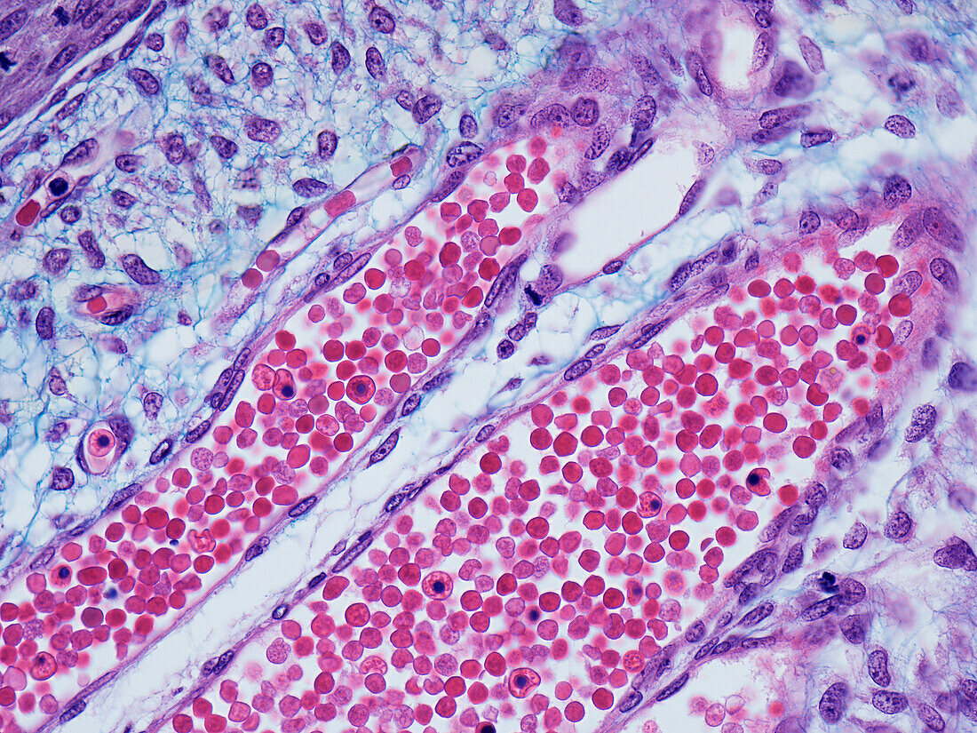
| Venule, light micrograph. Longitudinal section showing venules filled with erythrocytes (red blood cells) and occasional leukocytes (white blood cells) with nuclei. Venules have thin walls comprised of endothelial cells with slender elongated nuclei. External to the endothelial wall are pericytes which are fibroblasts and mesenchymal cells. At upper left several small calibre capillaries are seen in longitudinal and cross-section just wide enough to accommodate erythrocytes seen within the vessel lumen. Paraffin section, Masson's trichrome stain. Magnification: x320 when width printed at 10cm. | |
| Lizenzart: | Lizenzpflichtig |
| Credit: | Science Photo Library / Microscape |
| Bildgröße: | 4843 px × 3632 px |
| Modell-Rechte: | nicht erforderlich |
| Eigentums-Rechte: | nicht erforderlich |
| Restrictions: | - |
Preise für dieses Bild ab 15 €
Universitäten & Organisationen
(Informationsmaterial Digital, Informationsmaterial Print, Lehrmaterial Digital etc.)
ab 15 €
Redaktionell
(Bücher, Bücher: Sach- und Fachliteratur, Digitale Medien (redaktionell) etc.)
ab 30 €
Werbung
(Anzeigen, Aussenwerbung, Digitale Medien, Fernsehwerbung, Karten, Werbemittel, Zeitschriften etc.)
ab 55 €
Handelsprodukte
(bedruckte Textilie, Kalender, Postkarte, Grußkarte, Verpackung etc.)
ab 75 €
Pauschalpreise
Rechtepakete für die unbeschränkte Bildnutzung in Print oder Online
ab 495 €